. 2010 Jul;6(5):614-21.
doi: 10.4161/auto.6.5.12189. Epub 2010 Jul 1.

Keap1 facilitates p62-mediated ubiquitin aggregate clearance via autophagy

Affiliations

Keap1 facilitates p62-mediated ubiquitin aggregate clearance via autophagy

Weiliang Fan et al. Autophagy. 2010 Jul.

Abstract

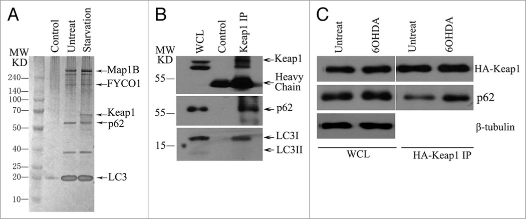
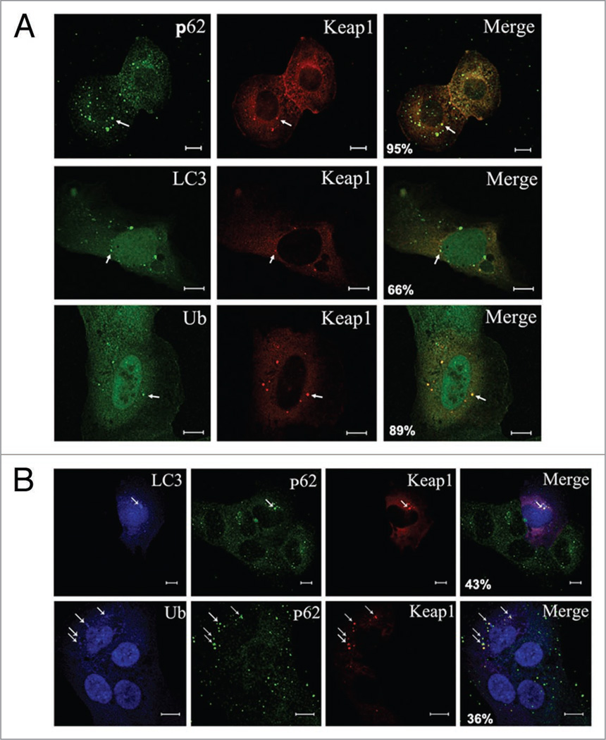
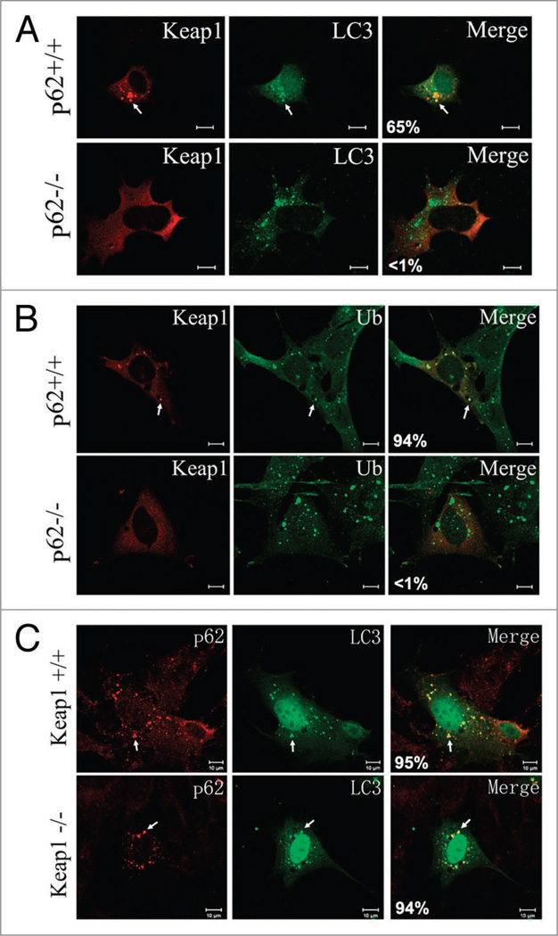
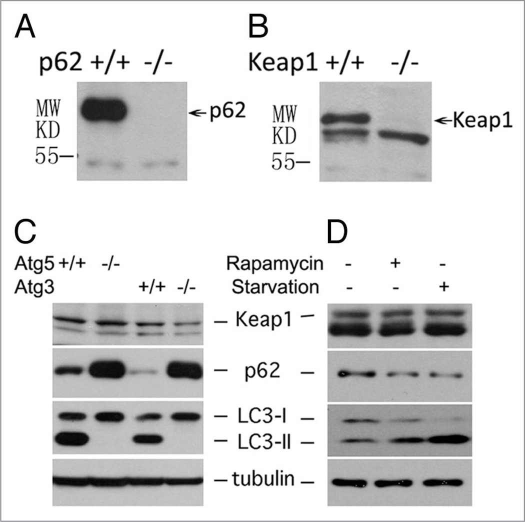
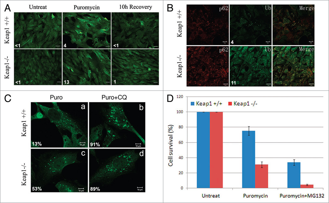
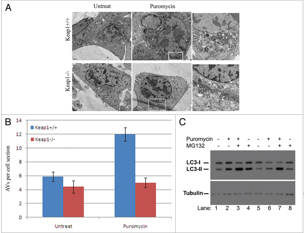
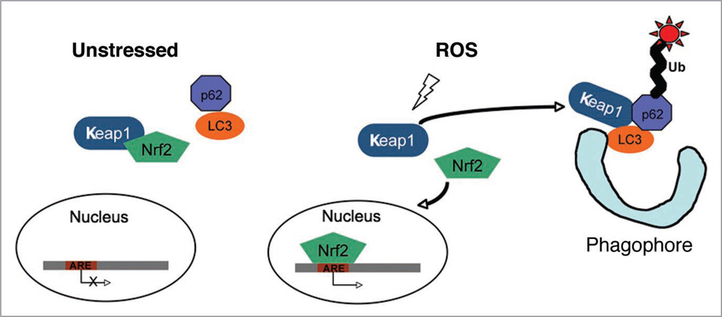
The accumulation of ubiquitin-positive protein aggregates has been implicated in the pathogenesis of neurodegenerative diseases, heart disease and diabetes. Emerging evidence indicates that the autophagy lysosomal pathway plays a critical role in the clearance of ubiquitin aggregates, a process that is mediated by the ubiquitin binding protein p62. In addition to binding ubiquitin, p62 also interacts with LC3 and transports ubiquitin conjugates to autophagosomes for degradation. The exact regulatory mechanism of this process is still largely unknown. Here we report the identification of Keap1 as a binding partner for p62 and LC3. Keap1 inhibits Nrf2 by sequestering it in the cytosol and preventing its translocation to the nucleus and activation of genes involved in the oxidative stress response. In this study, we found that Keap1 interacts with p62 and LC3 in a stress-inducible manner, and that Keap1 colocalizes with LC3 and p62 in puromycin-induced ubiquitin aggregates. Moreover, p62 serves as a bridge between Keap1 and ubiquitin aggregates and autophagosomes. Finally, genetic ablation of Keap1 leads to the accumulation of ubiquitin aggregates, increased cytotoxicity of misfolded protein aggregates, and defective activation of autophagy. Therefore, this study assigns a novel positive role of Keap1 in upregulating p62-mediated autophagic clearance of ubiquitin aggregates.


Figures

Figure 1
Figure 1
Keap1 interacts with LC3 and p62 in a stress-inducible manner. (A) Silver staining of LC3 complex after tandem affinity purification, LC3 complex was purified from U2OS cell that stably expresses ZZ-FLAG-LC3 with or without starvation for one hour. Proteins’ ID were identified by mass spectrometry. (B) Keap1 interacts with LC3 and p62 in vivo. HE K293T cells were immunoprecipitated with anti-Keap1 antibody, then immunoblotted with anti-Keap1 or anti-LC3 or anti-p62 antibodies. (C) 6-OHDA induces Keap1-p62 interaction. HA-Keap1 transfected HE K293T cells were treated with 6-OHDA for 6 hours. Cells were then lysed and followed with HA immunoprecipitation. The endogenous p62 pulled down by Keap1 was detected by western blotting. These experiments have been repeated at least three times.
Figure 2
Figure 2
Keap1 colocalizes with LC3 and p62 in the ubiquitin aggregates. (A) Keap1 colocalizes with p62, LC3 and ubiquitin in the puromycin-treated cells. U2OS cells were transfected with either DsRED-Keap1 or DsRED-Keap1 together with Myc-LC3. 2 days after transfection, cells were then fixed and stained with antibodies again p62, Myc and FK2 (for poly-ubiquitin chain) respectively. Scale bar 10 µm. Representative puncta are marked by arrows. The percentage of Keap1 puncta that colocalize with p62, LC3 or ubiquitin respectively was shown in the merged images. These data were summarized from at least 200 cells in three different experiments. (B) Coexistence of Keap1, LC3 and p62 in the ubiquitin-positive cellular puncta. U2OS cells were transfected with DsRED-Keap1 and Myc-LC3, followed by treatment with puromycin (10 µg/ml for 4 hours), cells were then fixed and p62 was stained with green fluorescent antibody, while Myc or ubiquitin were stained with blue AMCA conjugated secondary antibody. Scale bar 10 µm. Representative puncta are marked by arrows. The percentage of Keap1 puncta that colocalize with p62 and LC3/ubiquitin was shown in the merged images. These data were summarized from at least 200 cells in three different experiments.
Figure 3
Figure 3
p62 is required for Keap1 localization to ubiquitin aggregates and autophagosomes. (A) Wild-type or p62 knockout cells were transfected with DsRED-Keap1 together with Myc-LC3, followed by treatment with rapamycin (2 µM for 4 hours), cells were then fixed and Myc-LC3 was stained with a FITC secondary antibody. Scale bar 10 µm. The images are representative of at least three independent experiments. The percentage of Keap1 puncta that colocalize with LC3 was shown in the merged images. These data were summarized from at least 200 cells in three different experiments. (B) Wild-type or p62 knockout cells were transfected with DsRED-Keap1, followed by treatment with puromycin (10 µg/ml for 4 hours), cells were then fixed and ubiquitin was stained with a FITC conjugated secondary antibody. Scale bar 10 µm. The percentage of Keap1 puncta that colocalize with ubiquitin was shown in the merged images. These data were summarized from at least 200 cells in three different experiments. (C) Wild-type or Keap1 knockout cells were transfected with Myc-LC3, followed by treatment with rapamycin (2 µM for 4 hours). p62 and Myc-LC3 were stained with Rhodamine Red and FITC conjugated secondary antibodies. Scale bar 10 µm. The percentage of p62 puncta that colocalize with LC3 was shown in the merged images. These data were summarized from at least 200 cells in three different experiments.
Figure 4
Figure 4
Keap1 is not an autophagic substrate. (A) p62 expression was examined in wild-type or p62 knock out cells. (B) Keap1 expression was examined in wild-type or Keap1 knockout cells. (C) The protein level of Keap1, p62, tubulin and LC3 in Atg5+/+ and Atg5−/−, or Atg3+/+ and Atg3−/− mouse embryonic fibroblasts were detected by western blotting. (D) HE K293T cells were treated with either 2 µM rapamycin for 4 hours or starved by EBSS for 2 hours. After treatment, cells were lysed in 1× SDS, and Keap1, p62, tubulin and LC3 levels were detected by western blotting. All data shown in (C) and (D) are representative of at least three independent experiments.
Figure 5
Figure 5
Keap1 ablation leads to ubiquitin aggregates accumulation and cytotoxicity. (A) Ubiquitin aggregates in Keap1 knockout cells. Keap1+/+ and Keap1−/− MEF cells were treated with 10 µg/ml puromycin for 4 hours, and recovered in normal DMEM for another 10 hours. Cell were then fixed and stained with FK2 antibody. These images are representative of at least three independent experiments. The numbers of ubiquitin puncta per cell were summarized from at least 200 cells in three different experiments. (B) Keap1 wild-type and knockout MEFs were treated with puromycin for 4 hours. p62 and ubiquitin (Ub) were stained by anti-p62 or FK2 antibodies respectively. The numbers of ubiquitin puncta per cell were summarized from at least 200 cells in three different experiments. (C) Keap1+/+ and Keap1−/− cells were either treated with 10 µg/ml puromycin for 4 hours, or with 10 µg/ml puromycin and 200 µM chloroquine for 4 hours. Cells were then fixed and stained with FK2 antibody. These images are representative of at least three independent experiments. The percentage of large aggregates (twice the size of smaller ones) in total aggregates was summarized from at least 200 cells. (D) Keap1 wild-type and knockout cells were mock treated or treated with puromycin (10 µg/ml) alone, or co-treated with puromycin and MG132 (2 µM) for 4 hours. Cell numbers were counted 2 days after treatments in a Trypan blue assay. There data were summarized from three different experiments.
Figure 6
Figure 6
Genetic ablation of Keap1 leads to defective autophagy activation. (A) Electron microscopy analysis of Keap1+/+ and Keap1−/− cells. Puromycin (10 µg/ml, 4 hours) treated and untreated cells were analyzed under transmission electron microscope. The right panel is the enlarged image in the white frame. Scale bar, regular image: 2 µm, enlarge image: 0.5 µm. These experiments have been repeated at least three times. (B) The number of autophagic vesicles per cell cross section was calculated and quantified in this chart. There data were summarized from at least three different experiments. (C) LC3 conjugation was examined in Keap1 wildtype (Lanes 1–4) and knockout (Lanes 5–8) upon treatment of puromycin in combination with MG132 by western blotting. These experiments have been repeated at least three times.
Figure 7
Figure 7
Proposed model of the function of Keap1 in oxidative stress response and autophagy. In the unstressed cells, Keap1 sequesters Nrf2 in the cytosol. Keap1 is oxidized by reactive oxygen species (ROS), and Keap1 oxidation leads to Nrf2 dissociation. Nrf2 translocates to the nucleus and activates antioxidant response element (ARE)-containing genes. ROS also promotes the formation of Keap1-p62-LC3 complex that delivers ubiquitinated protein aggregates to the forming autophagosomes (phagophore).

Similar articles

Cited by

References

    1. Rubinsztein DC, Gestwicki JE, Murphy LO, Klionsky DJ. Potential therapeutic applications of autophagy. Nat Rev Drug Discov. 2007;6:304–312. - PubMed
    1. Mizushima N, Levine B, Cuervo AM, Klionsky DJ. Autophagy fights disease through cellular self-digestion. Nature. 2008;451:1069–1075. - PMC - PubMed
    1. Shintani T, Klionsky DJ. Autophagy in health and disease: a double-edged sword. Science. 2004;306:990–995. - PMC - PubMed
    1. Levine B, Kroemer G. Autophagy in the pathogenesis of disease. Cell. 2008;132:27–42. - PMC - PubMed
    1. Hara T, Nakamura K, Matsui M, Yamamoto A, Nakahara Y, Suzuki-Migishima R, et al. Suppression of basal autophagy in neural cells causes neurodegenerative disease in mice. Nature. 2006;441:885–889. - PubMed

Publication types

MeSH terms

LinkOut - more resources