

Comparison of optical coherence tomography and scanning laser polarimetry measurements in patients with multiple sclerosis
- PMID: 20495500
- PMCID: PMC2928137
- DOI: 10.1097/OPX.0b013e3181e3dcb3
Comparison of optical coherence tomography and scanning laser polarimetry measurements in patients with multiple sclerosis
Abstract
Purpose: To compare optical coherence tomography (OCT) and scanning laser polarimetry (GDx) measurements of the retinal nerve fiber layer (RNFL) in multiple sclerosis (MS) patients with and without optic neuritis (ON).
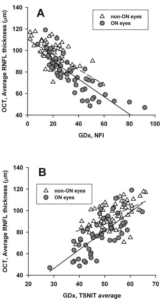
Methods: OCT and GDx were performed on 68 MS patients. Qualifying eyes were divided into two groups: 51 eyes with an ON history > or =6 months before (ON eyes) and 65 eyes with no history of ON (non-ON eyes). Several GDx and OCT parameters and criteria were used to define an eye as abnormal, for example, GDx nerve fiber indicator (NFI) >20 or 30, OCT average RNFL thickness, and GDx temporal-superior-nasal-inferior-temporal average (TSNIT) below 5 or 1% of the normative database of the instruments. Agreement between OCT and GDx parameters was reported as percent of observed agreement, along with the AC1 statistic. Linear regression analyses were used to examine the relationship between OCT average RNFL thickness and GDx NFI and TSNIT.
Results: All OCT and GDx measurements showed significantly more RNFL damage in ON than in non-ON eyes. Agreement between OCT and GDx parameters ranged from 69 to 90% (AC1 0.37 to 0.81) in ON eyes and 52 to 91% (AC1 = 0.21 to 0.90) in non-ON eyes. Best agreement was observed between OCT average RNFL thickness (p < 0.01) and NFI (>30) in ON eyes (90%, AC1 = 0.81) and between OCT average RNFL thickness (p < 0.01) and GDx TSNIT average (p < 0.01) in non-ON eyes (91%, AC1 = 0.90). In ON eyes, the OCT average RNFL thickness showed good linear correlation with NFI (R = 0.69, p < 0.0001) and TSNIT (R = 0.55, p < 0.0001).
Conclusions: OCT and GDx show good agreement and can be useful in detecting RNFL loss in MS/ON eyes.
Conflict of interest statement
The authors indicate no financial conflict of interest.
Figures
Similar articles
-
Retinal imaging by laser polarimetry and optical coherence tomography evidence of axonal degeneration in multiple sclerosis.Arch Neurol. 2008 Jul;65(7):924-8. doi: 10.1001/archneur.65.7.924. Arch Neurol. 2008. PMID: 18625859
-
Retinal nerve fiber layer thickness in subgroups of multiple sclerosis, measured by optical coherence tomography and scanning laser polarimetry.J Neurol. 2010 Oct;257(10):1654-60. doi: 10.1007/s00415-010-5589-1. Epub 2010 May 12. J Neurol. 2010. PMID: 20461397 Free PMC article.
-
Retinal nerve fiber layer in primary open-angle glaucoma with high myopia determined by optical coherence tomography and scanning laser polarimetry.Chin Med J (Engl). 2013;126(8):1425-9. Chin Med J (Engl). 2013. PMID: 23595371
-
Comparison of optical coherence tomography and scanning laser polarimetry for detection of localized retinal nerve fiber layer defects.J Glaucoma. 2010 Apr-May;19(4):229-36. doi: 10.1097/IJG.0b013e3181b21e87. J Glaucoma. 2010. PMID: 19730122
-
Scanning laser polarimetry and optical coherence tomography for detection of retinal nerve fiber layer defects.Korean J Ophthalmol. 2009 Sep;23(3):169-75. doi: 10.3341/kjo.2009.23.3.169. Epub 2009 Sep 8. Korean J Ophthalmol. 2009. PMID: 19794943 Free PMC article.
Cited by
-
Comparative study of the retinal nerve fibre layer thickness performed with optical coherence tomography and GDx scanning laser polarimetry in patients with primary open-angle glaucoma.Med Sci Monit. 2012 Mar;18(3):CR195-9. doi: 10.12659/msm.882525. Med Sci Monit. 2012. PMID: 22367131 Free PMC article.
-
Optical coherence tomography in multiple sclerosis and neuromyelitis optica: an update.Mult Scler Int. 2011;2011:472790. doi: 10.1155/2011/472790. Epub 2011 Jun 2. Mult Scler Int. 2011. PMID: 22096638 Free PMC article.
References
-
- Cantore WA. Optic neuritis. Pa Med. 1996;99 Suppl:96–98. - PubMed
-
- Sorensen TL, Frederiksen JL, Bronnum-Hansen H, Petersen HC. Optic neuritis as onset manifestation of multiple sclerosis: a nationwide, long-term survey. Neurology. 1999;53:473–478. - PubMed
-
- Frisen L, Hoyt WF. Insidious atrophy of retinal nerve fibers in multiple sclerosis. Funduscopic identification in patients with and without visual complaints. Arch Ophthalmol. 1974;92:91–97. - PubMed
-
- Elbol P, Work K. Retinal nerve fiber layer in multiple sclerosis. Acta Ophthalmol (Copenh) 1990;68:481–486. - PubMed
-
- Evangelou N, Konz D, Esiri MM, Smith S, Palace J, Matthews PM. Size-selective neuronal changes in the anterior optic pathways suggest a differential susceptibility to injury in multiple sclerosis. Brain. 2001;124:1813–1820. - PubMed
Publication types
MeSH terms
Grants and funding
LinkOut - more resources
Full Text Sources
Medical
Miscellaneous
