. 2010 Jul 7;29(13):2114-25.
doi: 10.1038/emboj.2010.100. Epub 2010 May 21.

Balanced ubiquitylation and deubiquitylation of Frizzled regulate cellular responsiveness to Wg/Wnt

Affiliations

Balanced ubiquitylation and deubiquitylation of Frizzled regulate cellular responsiveness to Wg/Wnt

Akiko Mukai et al. EMBO J. .

Abstract

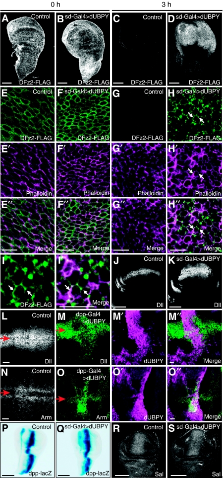
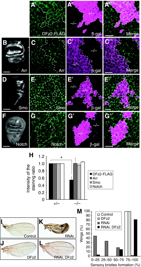
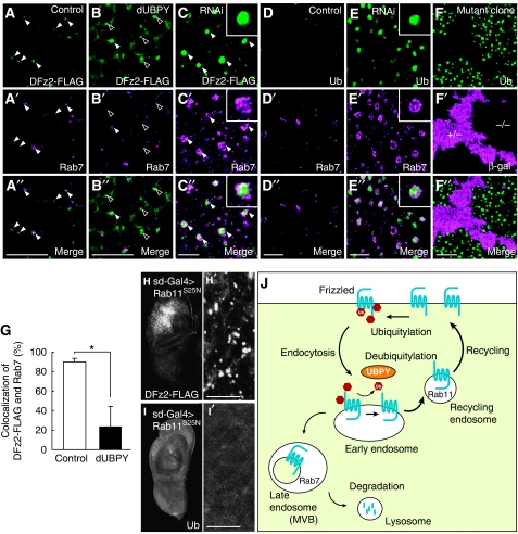
Wingless (Wg)/Wnt has been proposed to exert various functions as a morphogen depending on the levels of its signalling. Therefore, not just the concentration of Wg/Wnt, but also the responsiveness of Wg/Wnt-target cells to the ligand, must have a crucial function in controlling cellular outputs. Here, we show that a balance of ubiquitylation and deubiquitylation of the Wg/Wnt receptor Frizzled determines the cellular responsiveness to Wg/Wnt both in mammalian cells and in Drosophila, and that the cell surface level of Frizzled is regulated by deubiquitylating enzyme UBPY/ubiquitin-specific protease 8 (USP8). Although ubiquitylated Frizzled underwent lysosomal trafficking and degradation, UBPY/USP8-dependent deubiquitylation led to recycling of Frizzled to the plasma membrane, thereby elevating its surface level. Importantly, a gain and loss of UBPY/USP8 function led to up- and down-regulation, respectively, of canonical Wg/Wnt signalling. These results unveil a novel mechanism that regulates the cellular responsiveness to Wg/Wnt by controlling the cell surface level of Frizzled.


Conflict of interest statement

The authors declare that they have no conflict of interest.

Figures

Figure 1
Figure 1
dUBPY regulates Wg signalling in the wing disc. (A) Domain structures of human hUBPY and Drosophila dUBPY. Positions of the Cys- and His-boxes, MIT domain, rhodanese homology (RH) domain, and SH3-binding motifs (SBMs), as well as the dsRNA-target sites used in this study, are indicated. (BE) Top panels show wings from (B) control (sd-Gal; UAS-GFP RNAi), (C) RNAi-1 (sd-Gal; UAS-lacZ/UAS-dUBPY RNAi-1), (D) RNAi-2 (sd-Gal4; UAS-lacZ/UAS-dUBPY RNAi-2), and (E) partially rescued knockout (UAS-dUBPY; UBPY K.O.) flies. Bottom panels show high-magnification images of the wing margins indicated by arrowheads in top panels. (FI) Activity staining of β-gal derived from neuralized-lacZ (A101) (F, G) and wg-lacZ (H, I) in control (F, H) and dUBPY RNAi (G, I) wing discs. Arrows indicate the positions of the dorso-ventral border (F, G). (JK″) Anti-Sens (J) and anti-Arm (K) staining of dUBPY knockout clones, which are negative for β-gal (J′, K′, −/−), in the wing disc. J″ and K″ are merged images. Bars, 100 μm (FI); 10 μm (J″, K″).
Figure 2
Figure 2
UBPY facilitates canonical Wnt signalling in mammalian cells. (A) HEK293T cells were transfected with control FOP-FLASH or TOP-FLASH luciferase together with the indicated FLAG-UBPY constructs, and treated with Wnt3a overnight. Relative luciferase activity in the cell lysates is shown as mean±s.d. (n=4, *P<0.02, t-test). (B) NIH3T3 cells were transfected with the indicated FLAG-UBPY constructs, treated with Wnt3a for 1.5 h, and the lysates were immunoblotted with indicated antibodies. Positions of phosphorylated (P-Dvl-2) and non-phosphorylated Dvl-2 are indicated (top). The ratios of P-Dvl-2 to Dvl-2, which are normalized to that in mock-transfected cells stimulated with Wnt3a, are indicated below the top panel. (C) HEK293T cells were transfected with the indicated FLAG-UBPY constructs, treated with Wnt3a overnight, and their lysates were separated into cytoplasmic and membrane fractions. Each fraction was immunoblotted with indicated antibodies. FLAG-UBPY was solely recovered in the membrane fraction as described earlier (Mizuno et al, 2005). (D) HeLa cells were transfected with or without UBPY siRNAs and treated with Wnt3a for 5 h in the presence of cycloheximide. Cell lysates were immunoblotted with indicated antibodies. Relative intensity of the β-catenin bands is indicated (C, D).
Figure 3
Figure 3
Fz4 undergoes ubiquitylation and deubiquitylation in mammalian cells. (A) Schematic structure of mouse Fz4. Positions of the 12 Lys residues facing the cytoplasm are indicated. (B) HeLa cells were transfected with FLAG-Fz4 with or without HA-UBPYC748A, and treated with Wnt3a for 15 min. FLAG-Fz4 was immunoprecipitated and immunoblotted with indicated antibodies. (C) HeLa cells were transfected with HA-Fz4 with the indicated FLAG-Ub constructs. HA-Fz4 was immunoprecipitated and immunoblotted with indicated antibodies. (D) HeLa cells were transfected with FLAG-Fz4 with or without UBPY siRNAs, and treated with Wnt3a for 1 h. FLAG-Fz4 was immunoprecipitated and immunoblotted with indicated antibodies. (E) HeLa cells were transfected with FLAG-tagged Fz4 or Fz4K0 with or without HA-UBPYC748A, and labelled with biotin on the cell surface. Biotinylated FLAG-Fz4 proteins were immunoprecipitated and immunoblotted with indicated antibodies. (F) HeLa cells were transfected with FLAG-Fz4 with HA-tagged UBPYWT or UBPYC748A, and treated with Wnt3a for 1 h. FLAG-Fz4 was immunoprecipitated and immunoblotted with indicated antibodies. Total lysates were also immunoblotted with indicated antibodies (BF, lower panels). Asterisks indicate the IgG heavy chain used for immunoprecipitation (B, C, E, F). (G) FLAG-UBPY proteins, expressed in COS-7 cells and immunopurified with anti-FLAG beads, were stained with Coomassie Brilliant Blue (CBB) after electrophoresis (left). HA-Fz4 was expressed in HeLa cells, immunoprecipitated with anti-HA antibody, and incubated with the purified UBPY proteins. The reaction mixtures were immunoblotted with indicated antibodies (right).
Figure 4
Figure 4
UBPY regulates degradation and cell surface level of Fz4 in mammalian cells. (A) HeLa cells were transfected with FLAG-Fz4, labelled with biotin on the cell surface, and treated with or without Wnt3a for 3 or 6 h. FLAG-Fz4 was immunoprecipitated and blotted with streptavidin and anti-FLAG antibody. Arrows, closed arrowheads, and open arrowheads indicate Fz proteins conjugated with zero, one, and two Ub molecules, respectively. (B) HeLa cells were transfected with FLAG-Fz4 with HA-tagged UBPYC748A or UBPYS680A, labelled with biotin, and chased for 6 h. FLAG-Fz4 was immunoprecipitated and blotted with streptavidin and anti-FLAG antibody. Total lysates were immunoblotted with anti-HA antibody. (CD″) HEK293T cells were transfected with Fz4, which was FLAG-tagged extracellularly, together with HA-tagged UBPYS680A (CC″) or UBPYC748A (DD″). Living cells were stained with anti-FLAG antibody (C, D). After fixation, cells were further stained with anti-HA antibody and TO-PRO-3 (C′, D′). Arrows indicate cells expressing UBPYS680A or UBPYC748A. Arrowheads indicate cells expressing no ectopic UBPY. C″ and D″ are merged images. Bars, 10 μm. (E, F) Anti-FLAG fluorescence intensity of FLAG-Fz4-expressing cells in the experiments in CC″ and DD″ was quantified and shown as mean±s.d. (n [field of view]=10–25, *P<0.01, t-test). (G) HeLa cells were transfected with FLAG-tagged Fz4 or Fz4K0, labelled with biotin, and chased for 6 h. FLAG-Fz4 proteins were immunoprecipitated and blotted with streptavidin and anti-FLAG antibody. The intensity of the biotinylated Fz4 bands was quantified, and the ratio of the intensity after 3 or 6 h of chase to that at 0 h is shown as mean±s.d. (n=5, *P<0.01, t-test) (A, B, bottom; G, right). (H) HEK293T cells were transfected with FOP-FLASH or TOP-FLASH luciferase together with the indicated Fz4 or UBPY constructs, and treated with Wnt3a overnight. Relative luciferase activity in the cell lysates is shown as mean±s.d. (n=3, *P<0.02, t-test). (I) HEK293T cells were transfected with TOP-FLASH luciferase together with the indicated Fz4 and UBPY constructs and treated with Wnt3a overnight. Relative luciferase activity in UBPYC748A- and UBPYS680A-expressing cells to that in UBPYWT-expressing cells is shown (mean±s.d.; n=3, *P<0.02, t-test).
Figure 5
Figure 5
dUBPY regulates cell surface DFz2 level and Wg signalling in the wing disc. (AH″) Staining of control (A, C, EE″, GG″) and dUBPY-overexpressing (B, D, FF″, HH″) DFz2-FLAG wing discs at the end of heat shock induction of DFz2-FLAG (A, B, EE″, FF″) and after a 3-h chase at 25°C (C, D, GG″, HH″) with anti-FLAG antibody (AH) and phalloidin (E′–H′). E″–H″ are merged images. All the dUBPY-overexpressing discs examined (∼30) exhibited the same phenotype. (I, I′) High-magnification images of (H) and (H″). Arrows indicate the plasma membrane (HH″, I, I′). (J, K) Anti-Dll staining of control (J) and dUBPY-overexpressing (K) wing discs. (LO″) Anti-Dll (L, MM″) and anti-Arm (N, OO″) staining of control (L, N) and dpp-Gal4-driven dUBPY-overexpressing (MM″, OO″) wing discs. dUBPY-overexpressing discs were co-stained with anti-dUBPY antibody (M′, O′). M″ and O″ are merged images. Arrows indicate the positions of the dorso-ventral border (LO). (PS) Activity staining of β-gal derived from dpp-lacZ (P, Q) and anti-Sal (R, S) staining in control (P, R) and dUBPY-overexpressing (Q, S) wing discs. Bars, 100 μm (AD, J, K, PS); 10 μm (E″–H″, I′, L, M″, N, O″).
Figure 6
Figure 6
dUBPY is a selective regulator for DFz2 in the wing disc. (AA″) Anti-FLAG staining (A) of a dUBPY knockout clone, which is negative for β-gal (A′, −/−), in the DFz2-FLAG wing pouch after a 2-h chase of heat shock-induced DFz2-FLAG. (BG″) Anti-Arr (B, C), anti-Smo (D, E), and anti-Notch (F, G) staining of the wild-type wing discs (B, D, F) and dUBPY knockout clones (C, E, G), which are negative for β-gal (C′, E′, G′, −/−), in the wing pouch. A″, C″, E″, and G″ are merged images. Arrows in B, D, and F indicate regions that were analysed in the dUBPY knockout discs in CC″, EE″, and GG″. Bars, 10 μm (A″, C″, E″, G″); 100 μm (B, D, F). (H) Anti-FLAG (DFz2), anti-Arr, anti-Smo, and anti-Notch fluorescence intensity in experiments in AG″ was quantified in the control and dUBPY knockout areas. Relative intensity in the knockout area (−/−) to that in the control area (+/−) is shown (mean±s.d., n=12 for each staining, *P<0.02, t-test). (IL) Control (I), DFz2-overexpressing (J), dUBPY RNAi (K), and dUBPY RNAi/DFz2-overexpressing (L) wings in adult flies. (M) Quantification of sensory bristle formation in wings in experiments in IL. Percentage of wings with a different degree of sensory bristle formation is shown (n=33–62).
Figure 7
Figure 7
dUBPY regulates endocytic DFz2 trafficking in the wing disc. (AE″) Control (AA″, DD″), dUBPY-overexpressing (BB″), and dUBPY RNAi (CC″, EE″) wing discs expressing DFz2-FLAG (AC″) or not (DE″) were stained with anti-FLAG (AC) or FK2 (D, E) antibody together with anti-Rab7 (A′–E′). Closed and open arrowheads indicate Rab7-positive and Rab7-negative endosomes, respectively (AC″). Insets in CC″ and EE″ show high-magnification images of typical Rab7-positive endosomes. (FF″) FK2 staining (F) of a dUBPY knockout clone in the wing disc, which is negative for β-gal (F′, −/−). A″–F″ are merged images. (G) Control and dUBPY-overexpressing wing discs expressing DFz2-FLAG were double-stained with anti-FLAG and anti-Rab7 antibodies as in AA″ and BB″. Percentage of FLAG/Rab7-double-positive endosomes among total FLAG-positive endosomes was then determined (mean±s.d., n [field of view]=5–7, *P<0.01, t-test). (HI′) Anti-FLAG (H, H′) and FK2 (I, I′) staining of DFz2-FLAG wing discs co-expressing Rab11S25N in the wing pouch. H′ and I′ show high-magnification images of the wing pouch regions in H and I. Bars, 10 μm. (J) A model for the regulation of the cell surface level of Fz.

Comment in

References

    1. Bengochea A, de Souza MM, Lefrançois L, Le Roux E, Galy O, Chemin I, Kim M, Wands JR, Trepo C, Hainaut P (2008) Common dysregulation of Wnt/Frizzled receptor elements in human hepatocellular carcinoma. Br J Cancer 99: 143–150 - PMC - PubMed
    1. Blitzer JT, Nusse R (2006) A critical role for endocytosis in Wnt signaling. BMC Cell Biol 7: 28. - PMC - PubMed
    1. Cadigan KM, Fish MP, Rulifson EJ, Nusse R (1998) Wingless repression of Drosophila frizzled 2 expression shapes the Wingless morphogen gradient in the wing. Cell 93: 767–777 - PubMed
    1. Campuzano S, Modolell J (1992) Patterning of the Drosophila nervous system: the achaete-scute gene complex. Trends Genet 8: 202–208 - PubMed
    1. Chen W, ten Berge D, Brown J, Ahn S, Hu LA, Miller WE, Caron MG, Barak LS, Nusse R, Lefkowitz RJ (2003) Dishevelled 2 recruits β-arrestin 2 to mediate Wnt5A-stimulated endocytosis of Frizzled 4. Science 301: 1391–1394 - PubMed

Publication types