

Intraductal papillary mucinous neoplasm of the biliary tract: a real disease?
- PMID: 20495637
- PMCID: PMC2799622
- DOI: 10.1111/j.1477-2574.2009.00122.x
Intraductal papillary mucinous neoplasm of the biliary tract: a real disease?
Abstract
Background: Despite increasing numbers of reports, biliary tract intraductal papillary mucinous neoplasm (BT-IPMN) is not yet recognized as a unique neoplasm. The aim of the present study was to define the presence of BT-IPMN in a large series of resected biliary neoplasms.
Methods: From May 1994 to December 2006, BT-IPMN cases were identified by reviewing pathology specimens of all resected cholangiocarcinomas and other biliary neoplasms when cystic, papillary or mucinous features were cited in pathology reports.
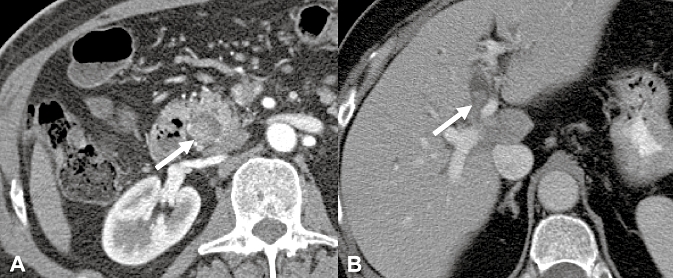
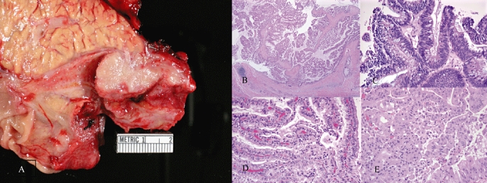
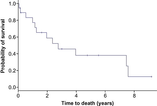
Results: BT-IPMN was identified in 23 out of 253 (9%) specimens using the strict histopathological criteria of IPMN. The most common presenting symptom was abdominal discomfort which was present in 15 patients (65%). Only one of the original operative pathology reports used the term IPMN; 16 (70%) used the terms cystic, mucinous and/or papillary. BT-IPMN was isolated to non-hilar extra-hepatic ducts in 12 (52%), intra-hepatic ducts in 6 (26%) and hilar extra-hepatic ducts in 5 patients (22%). Carcinoma was found in association with BT-IPMN in 19 patients (83%); 5-year survival was 38% after resection.
Conclusion: BT-IPMN occurs throughout the intra- and extra-hepatic biliary system and can be identified readily as a unique neoplasm. Broader acceptance of BT-IPMN as a unique neoplasm may lead to a better understanding of the pathogenesis of biliary malignancies.
Figures
Similar articles
-
Biliary tract intraductal papillary mucinous neoplasm: report of 19 cases.World J Gastroenterol. 2015 Apr 14;21(14):4261-7. doi: 10.3748/wjg.v21.i14.4261. World J Gastroenterol. 2015. PMID: 25892877 Free PMC article.
-
Magnetic Resonance Imaging versus Computed Tomography for Biliary Tract Intraductal Papillary Mucinous Neoplasm (BT-IPMN): A Diagnostic Performance Analysis.Med Sci Monit. 2020 Apr 1;26:e920952. doi: 10.12659/MSM.920952. Med Sci Monit. 2020. PMID: 32235820 Free PMC article.
-
A comparison between intraductal papillary neoplasms of the biliary tract (BT-IPMNs) and intraductal papillary mucinous neoplasms of the pancreas (P-IPMNs) reveals distinct clinical manifestations and outcomes.Eur J Surg Oncol. 2013 Jun;39(6):554-8. doi: 10.1016/j.ejso.2013.02.016. Epub 2013 Mar 15. Eur J Surg Oncol. 2013. PMID: 23506840
-
MRI features of intraductal papillary mucinous neoplasm of the bile ducts, "The myth about the cyst": A systematic review.Eur J Radiol Open. 2023 Aug 12;11:100515. doi: 10.1016/j.ejro.2023.100515. eCollection 2023 Dec. Eur J Radiol Open. 2023. PMID: 37609049 Free PMC article. Review.
-
Intraductal papillary mucininous neoplasm of the bile ducts: multimodality assessment with pathologic correlation.Abdom Imaging. 2011 Aug;36(4):447-56. doi: 10.1007/s00261-010-9649-x. Abdom Imaging. 2011. PMID: 20959978 Review.
Cited by
-
Intraductal Papillary Neoplasms of the Bile Duct: Clinical Case Insights and Literature Review.Clin Pract. 2024 Aug 27;14(5):1669-1681. doi: 10.3390/clinpract14050133. Clin Pract. 2024. PMID: 39311283 Free PMC article.
-
A huge intraductal papillary neoplasm of the bile duct treated by right trisectionectomy after right portal vein embolization.Ann Hepatobiliary Pancreat Surg. 2018 May;22(2):150-155. doi: 10.14701/ahbps.2018.22.2.150. Epub 2018 May 30. Ann Hepatobiliary Pancreat Surg. 2018. PMID: 29896576 Free PMC article.
-
Intraductal papillary neoplasm of the bile duct, gastric type, arising in the intrapancreatic common bile duct could progress to colloid carcinoma: report of a case.Int J Clin Exp Pathol. 2015 May 1;8(5):5848-55. eCollection 2015. Int J Clin Exp Pathol. 2015. PMID: 26191308 Free PMC article.
-
Intraductal papillary mucinous neoplasm of the liver: GI image.J Gastrointest Surg. 2015 Apr;19(4):792-4. doi: 10.1007/s11605-015-2750-2. Epub 2015 Jan 24. J Gastrointest Surg. 2015. PMID: 25617079
-
Intraductal papillary neoplasm of the bile duct: a single-center retrospective study.J Int Med Res. 2018 Oct;46(10):4258-4268. doi: 10.1177/0300060518792800. Epub 2018 Aug 15. J Int Med Res. 2018. PMID: 30111208 Free PMC article.
References
-
- Helpap B. Malignant papillomatosis of the intrahepatic bile ducts. Acta Hepatogastroenterol (Stuttg) 1977;24:419–425. - PubMed
-
- Neumann RD, LiVolsi VA, Rosenthal NS, Burrell M, Ball TJ. Adenocarcinoma in biliary papillomatosis. Gastroenterology. 1976;70(5):779–782. Pt. 1. - PubMed
-
- Kokubo T, Itai Y, Ohtomo K, Itoh K, Kawauchi N, Minami M. Mucin-hypersecreting intrahepatic biliary neoplasms. Radiology. 1988;168:609–614. - PubMed
-
- Cattell RB, Braasch JW, Kahn F. Polypoid epithelial tumors of the bile ducts. N Engl J Med. 1962;266:57–61. - PubMed
LinkOut - more resources
Full Text Sources
