

Giant paravertebral myxoma
- PMID: 20495934
- PMCID: PMC3111513
- DOI: 10.1007/s00586-010-1442-6
Giant paravertebral myxoma
Abstract
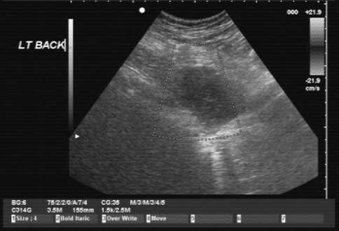
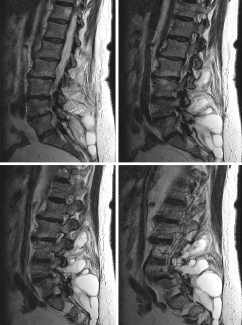
The study design includes case report and clinical discussion. The objective was to describe a rare case of a giant intramuscular myxoma (IMM) presenting as a mass in the paravertebral muscles. Myxoma is a rare benign soft tissue tumour of mesenchymal origin. Although intramuscular presentation is common, they are rare in the paravertebral muscles and are characteristically <5 cm in length. We report the clinical and imaging features in a 70-year-old woman presenting with back pain, asymmetry of the waist and a mass in right paravertebral region. This was originally misdiagnosed as a juxtafacet synovial cyst after CT-guided biopsy. The mass was excised en bloc and sent for histology. This revealed a low-grade myxoid neoplasm with features of an IMM. The patient went on to make a complete recovery. To our knowledge, this is only the fifth case of paravertebral IMM reported in the literature and at approximately 15 cm in length may be the largest encountered in clinical practice.
Figures


Similar articles
-
Intramuscular myxoma: characteristic MR imaging features.AJR Am J Roentgenol. 2002 May;178(5):1255-9. doi: 10.2214/ajr.178.5.1781255. AJR Am J Roentgenol. 2002. PMID: 11959742
-
Intramuscular myxoma of the cervical paraspinal muscle.Eur Spine J. 2009 Jul;18 Suppl 2(Suppl 2):245-9. doi: 10.1007/s00586-009-0933-9. Epub 2009 Mar 20. Eur Spine J. 2009. PMID: 19301043 Free PMC article. Review.
-
Sonographic appearance of an intramuscular myxoma of the pectoralis major muscle.J Clin Ultrasound. 2014 Oct;42(8):505-8. doi: 10.1002/jcu.22149. Epub 2014 Mar 13. J Clin Ultrasound. 2014. PMID: 24633968
-
Intramuscular myxoma with fibrous dysplasia: a report of two cases with a review of the literature.Pathol Int. 1995 Feb;45(2):165-71. doi: 10.1111/j.1440-1827.1995.tb03438.x. Pathol Int. 1995. PMID: 7742929 Review.
-
A case of intramuscular lumbar myxoma: Uncertainty in the preoperative diagnosis of a spinal soft tissue tumour.Neurochirurgie. 2022 Oct;68(5):530-534. doi: 10.1016/j.neuchi.2021.08.007. Epub 2021 Sep 16. Neurochirurgie. 2022. PMID: 34537211
Cited by
-
Intramuscular myxoma of the buttock mimicking low-grade fibromyxoid sarcoma: diagnostic usefulness of MUC4 expression.Skeletal Radiol. 2013 Oct;42(10):1475-9. doi: 10.1007/s00256-013-1641-4. Epub 2013 May 19. Skeletal Radiol. 2013. PMID: 23685711
-
Lumbar Intramuscular Myxoma: Microsurgical Resection With Assistance From an Endoscopic Microinspection Tool.Int J Spine Surg. 2025 Jun 12;19(3):324-329. doi: 10.14444/8733. Int J Spine Surg. 2025. PMID: 40174973 Free PMC article.
-
Lumbar paraspinal intramuscular myxoma: A case report.Surg Neurol Int. 2023 Mar 10;14:86. doi: 10.25259/SNI_141_2023. eCollection 2023. Surg Neurol Int. 2023. PMID: 37025538 Free PMC article.
-
Cellular myxoma of the lumbar spine.Surg Neurol Int. 2013 Jun 19;4:82. doi: 10.4103/2152-7806.113648. Print 2013. Surg Neurol Int. 2013. PMID: 23869282 Free PMC article.
-
Atypical Intramuscular Myxoma of the Lumbosacral Paraspinal Muscle: The First Case Report in Asian.J Korean Neurosurg Soc. 2015 Dec;58(6):566-70. doi: 10.3340/jkns.2015.58.6.566. Epub 2015 Dec 31. J Korean Neurosurg Soc. 2015. PMID: 26819696 Free PMC article.
References
-
- Caro P, Dubrana F, Le Nen D, et al. Myxome intramusculaire. Rev Chir Orthop. 1991;77:568–570. - PubMed
-
- Heymans O, Gebhart M, Alexiou J, et al. Intramuscular myxoma. Acta Chir Belg. 1998;98:120–122. - PubMed
-
- Enzinger FM. Intramuscular myxoma, a review and follow-up study of 34 cases. Am J Clin Pathol. 1965;43:104–113. - PubMed
-
- Bancroft LW, Kransdorf MJ, Menke DM, O’Connor MI, Foster WC. Intramuscular myxoma: characteristic MR imaging features. AM J Roentenol. 2002;178(5):1255–1259. - PubMed
Publication types
MeSH terms
LinkOut - more resources
Full Text Sources
Research Materials
