. 2010 Jul;28(7):1231-42.
doi: 10.1002/stem.449.

EphB3 limits the expansion of neural progenitor cells in the subventricular zone by regulating p53 during homeostasis and following traumatic brain injury

Affiliations

EphB3 limits the expansion of neural progenitor cells in the subventricular zone by regulating p53 during homeostasis and following traumatic brain injury

Michelle H Theus et al. Stem Cells. 2010 Jul.

Abstract

Ephrins and Eph receptor(s) have recently been implicated in regulating neurogenesis in the adult subventricular zone (SVZ) and rostral migratory stream. Here, we examined the role of ephrinB3-EphB3 signaling in mediating the SVZ response to traumatic brain injury (TBI). Analysis of EphB3 expression showed colocalization with glial fibrillary acidic protein-positive neural stem progenitor cells (NSPCs) and doublecortin-positive neuroblasts, whereas ephrinB3 was expressed outside the neurogenic region. TBI resulted in a significant reduction in EphB3 expression, which coincided with enhanced NSPC survival and proliferation at 3 and 7 days postinjury. Analysis of mice lacking either ephrinB3 (ephrinB3(-/-)) or EphB3 (EphB3(-/-)) showed a significant increase in bromodeoxyuridine (BrdU) incorporation and Ki67 immunoreactivity in the SVZ. Interestingly, cell death was dissimilar between knockout mice, where cell death was reduced in EphB3(-/-) but increased in ephrinB3(-/-) mice. Lateral ventricle infusion of soluble preclustered ephrinB3-Fc reversed the proliferative and cell death defects in ephrinB3(-/-) but not EphB3(-/-) mice and prevented TBI-induced proliferation in wild-type NSPCs. Coincidently, tumor suppressor p53 expression was increased following EphB3 stimulation and is reduced in the absence of either EphB3 or ephrinB3. Furthermore, pharmacological inhibition and siRNA knockdown of p53-attenuated ephrinB3-Fc-mediated growth suppression while having no effect on cell death in cultured NSPCs. These data demonstrate that EphB3 signaling suppresses NSPC proliferation in a p53-dependent manner, induces cell death in the absence of ligand stimulation and is transiently reduced in the SVZ to initiate the expansion and survival of endogenous adult NSPCs following TBI.


Figures
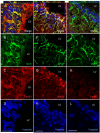
Figure 1
Figure 1
EphB3 is expressed on GFAP-positive NSPCs and DCX-positive neuroblasts in the adult SVZ. (A-D) Confocal z-stack image analysis of EphB3 (red) using double-immunofluorescence showed co-labeling with doublecortin (DCX)-positive cells (green) in the lateral wall of the lateral ventricle. (E-H) GFAP-positive cells (green) also co-label with anti-EphB3 (red) in cytoplasmic extension in the adult SVZ. (I-L) Sagittal tissue sections from EphB3−/− mice do not show expression with anti-EphB3 antibody (red) and does not co-label with GFAP-expressing cells. Scale bar = 20 μm. LV, lateral ventricle.
Figure 2
Figure 2
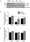
EphB3 expression is down-regulated in the SVZ following CCI injury. (A) Western blot analysis of EphB3 and ephrinB3 from the ipsilateral (i) and contralateral (c) SVZ in sham and CCI-injured wild type mice at 1, 3 and 7 days. EphB3 expression is significantly reduced at both 3 and 7 days but not 1 day after CCI injury. EphrinB3 expression was unchanged in the first 7 days after injury. Quantified (B) EphB3 and (C) ephrinB3 expression in the SVZ of CCI-injured mice (n=5) compared to sham-injured mice (n=5) at 1, 3 and 7 days. Quantified data was normalized to β-actin control levels then represented as relative expression compared to sham-injury at each time point. *P<0.05; ***P<0.001 compared to ipsilateral sham-injured mice. #P<0.05; ##P<0.01 compared to contralateral sham-injured mice. KO CTL=whole brain extract from EphB3−/− mice.
Figure 3
Figure 3
Proliferation in the adult SVZ after CCI injury in wild type, ephrinB3−/− and EphB3−/− mice. (A) Stereological counts of Ki67-positive cells at 1, 3 and 7 days post-injury. CCI increased proliferation at 3 and 7 days in wild type mice while having no effect in ephrinB3−/− and EphB3−/− ipsilateral SVZ. Greater numbers of Ki67-positve cells are found in sham- and CCI-injured (1 and 3 days) ephrinB3−/− and EphB3−/− mice as compared to wild type mice. (B) BrdU-incorporation at 3 days confirms the increase in proliferation in CCI-injured wild type ipsilateral SVZ, while loss of ephrinB3 and EphB3 enhances proliferation in both sham and CCI-injured mice. (C) CCI-induced proliferation is reversed in wild type and ephrinB3−/− but not EphB3−/− mice following 3 days infusion of pre-clustered soluble ephrinB3-Fc (eB3-Fc) compared to Fc-control infusion. (D-F) Confocal image analysis of BrdU (red) and PSA-NCAM (green) immunofluorescence from sagittal sections through the SVZ and RMS of wild type (D1-D3), ephrinB3−/− (E1-E3) and EphB3−/− (F1-F3). Co-expression of BrdU-positive cells is observed in PSA-NCAM-positive neuroblasts in the SVZ and greater PSA-NCAM staining is observed in ephrinB3−/− and EphB3−/− mice. (High-magnification images, scale bar = 20 μm; Low magnification: Scale bar = 100 μm). *P<0.05, **P<0.01, ***P<0.001 compared to wild type sham. #P<0.05, ###P<0.001 compared to corresponding wild type CCI. LV, lateral ventricle.
Figure 4
Figure 4
EphB3 regulates cell death in the SVZ of sham and CCI-injured mice. (A) Stereological counts of TUNEL-positive cells in the adult SVZ of wild type, ephrinB3−/− and EphB3−/− mice. The number of TUNEL-positive cells is reduced in the ipsilateral SVZ 3 days after CCI injury in wild type and ephrinB3−/−; but remains significantly lower in both sham- and CCI-injured EphB3−/− mice. Interestingly, there is greater cell death in the absence of eprhinB3 but reduced cell death in the absence of EphB3 compared to wild type in sham-injured animals. (B) Infusion of pre-clustered eB3-Fc can restore the level of cell death in ephrinB3−/− mice to wild type levels and significantly reduce the basal level of cell death in sham-injured wild type mice. The number of TUNEL-positive cells in the SVZ of EphB3−/− mice is unaffected by eB3-Fc infusion nor is there any effect in CCI-injured SVZ tissue. *P<0.05, ***P<0.001 compared to wild type sham; ###P<0.001 compared to ephrinB3−/− sham in panel A and *P<0.05 compared to corresponding Fc-control in panel B.
Figure 5
Figure 5
Tumor suppressor p53 expression is down-regulated in the wild type adult SVZ following brain injury and is expressed at lower levels in ephrinB3−/− and EphB3−/− mice. (A-D) Confocal images of p53 immunofluorescence in the SVZ of sagittal wild type tissue sections. (A’-D’) Confocal image analysis at 100X magnification. Co-localization of p53 (green) and EphB3 (red) is observed in cytoplasmic processes (yellow arrow). Nuclear staining of p53 is also seen in EphB3-expressing cells in the SVZ (white arrowheads). (E-H) Confocal images of p53 immunofluorescence in the SVZ of p53−/− mice. EphB3 (red) is expressed in the SVZ, while p53 staining is not observed in p53-null mice. (I) Western blot analysis of p53 expression 3 days after sham and CCI injury in wild type, EphB3−/− and ephrinB3−/− mice. P53 is reduced in the ipsilateral (i) and contralateral (c) SVZ after CCI injury, while lower levels of p53 remain in EphB3−/− and ephrinB3−/− mice. (J) Bar graph represents quantified data of p53 in the ipsilateral SVZ in sham- and CCI-injured wild type and knockout mice. Data represents relative p53 expression normalized to β-actin control levels. (n=4). *P<0.05; **P<0.01 compared to wild type sham. LV, lateral ventricle. (A-D and E-H) Scale bar = 20 μm, (A’-D’) Scale bar = 10 μm.
Figure 6
Figure 6
EphB3 directly regulates p53 and AKT phosphorylation in the SVZ. EphB3 was stimulated in vivo by infusing pre-clustered ephirnB3-Fc (eB3-Fc) or Fc-control (Fc) molecules into the lateral ventricle of wild type and EphB3−/− mice. (A) Western blot analysis of p53 protein expression 3 days after eB3-Fc and Fc infusions show an increase in p53 expression in the ipsilateral (i) and contralateral (c) SVZ of wild type but not EphB3−/− mice. (B) Bar graph representing quantified data of p53 normalized to β-actin control levels in the SVZ of wild type and EphB3−/− mice (n=4). (C) The level of phosphorylated AKT (p-AKT) is decreased in the ipsilateral SVZ 3 days after eB3-Fc stimulation as total p53 and phosphoSer15-p53 are increased in wild type while having no effect in EphB3−/− mice. (D) Bar graph representing quantified data of p-AKT levels normalized to total AKT levels in the SVZ of wild type and EphB3−/− mice (n=4). *P<0.05 and **P<0.01 compared to wild type Fc-control.
Figure 7
Figure 7
EphB3 regulates proliferation of cultured SVZ-derived adult NSPCs in a p53-dependent manner. (A-D) Triple immunofluorescence labeling of BrdU-positive (red) wild type NSPCs with nestin (green) NSPC marker and GFAP (purple). EphB3−/− NSPCs have more proliferating nestin-expressing, GFAP-positive cells compared to wild type cultures. (I) Proliferation of wild type and EphB3−/− NSPCs in the presence and absence of soluble pre-clustered ephrinB3-Fc (eB3-Fc) and Fc-control (Fc). The percentage of BrdU-positive cells is increased in EphB3−/− cultures. The addition of 5 μg/ml eB3-Fc molecules for 3 days suppressed proliferation in wild type NSPCs compared to Fc-control, while having no effect in EphB3−/− cells. (J) The percentage of basal level cell death is reduced in EphB3−/− compared to wild type NSPCs. The addition of either 1 μg/ml or 5 μg/ml eB3-Fc reduced the percentage of cell death in wild type NSPCs compared to Fc-control, while having no effect in EphB3−/− cells. (K) EB3-Fc induced growth suppression (white bars) is attenuated in the presence of 10μM p53 inhibitor, Pifithrin compared to PBS-vehicle control. (L) Pre-treatment with si p53, 24 hours prior to stimulation also attenuated eB3-Fc induced growth suppression compared to siRNA scramble control. (M) EphB3 stimulation induces nuclear translocation of total p53 and phosphoSer15-p53 following 15 min eB3-Fc exposures compared to Fc-control (c). Inhibiting p53 with 10 μM Pifithrin (N) or si p53 (O) had no effect on altering the percentage of cell death in the presence of 5 μg/ml ephrinB3-Fc. (**P<0.01, ***P<0.001 compared to corresponding wild type Fc-control; ###P<0.001 compared to wild type control in panel I and J), (*P<0.05 compared to Fc-control, ##P<0.01 compared to eB3-Fc plus vehicle in panel K, Scale bar = 200 μM).

Similar articles

Cited by

References

    1. Gage FH. Mammalian neural stem cells. Science. 2000;287(5457):1433–8. - PubMed
    1. Sohur US, et al. Adult neurogenesis and cellular brain repair with neural progenitors, precursors and stem cells. Philos Trans R Soc Lond B Biol Sci. 2006;361(1473):1477–97. - PMC - PubMed
    1. Kempermann G, Gage FH. New nerve cells for the adult brain. Sci Am. 1999;280(5):48–53. - PubMed
    1. Eriksson PS, et al. Neurogenesis in the adult human hippocampus. Nat Med. 1998;4(11):1313–7. - PubMed
    1. Altman J, Das GD. Autoradiographic and histological studies of postnatal neurogenesis. I. A longitudinal investigation of the kinetics, migration and transformation of cells incorporating tritiated thymidine in neonate rats, with special reference to postnatal neurogenesis in some brain regions. J Comp Neurol. 1966;126(3):337–89. - PubMed

Publication types

MeSH terms