

Histone deacetylases are critical regulators of the renin-angiotensin system during ureteric bud branching morphogenesis
- PMID: 20496471
- PMCID: PMC3039915
- DOI: 10.1203/PDR.0b013e3181da477c
Histone deacetylases are critical regulators of the renin-angiotensin system during ureteric bud branching morphogenesis
Abstract
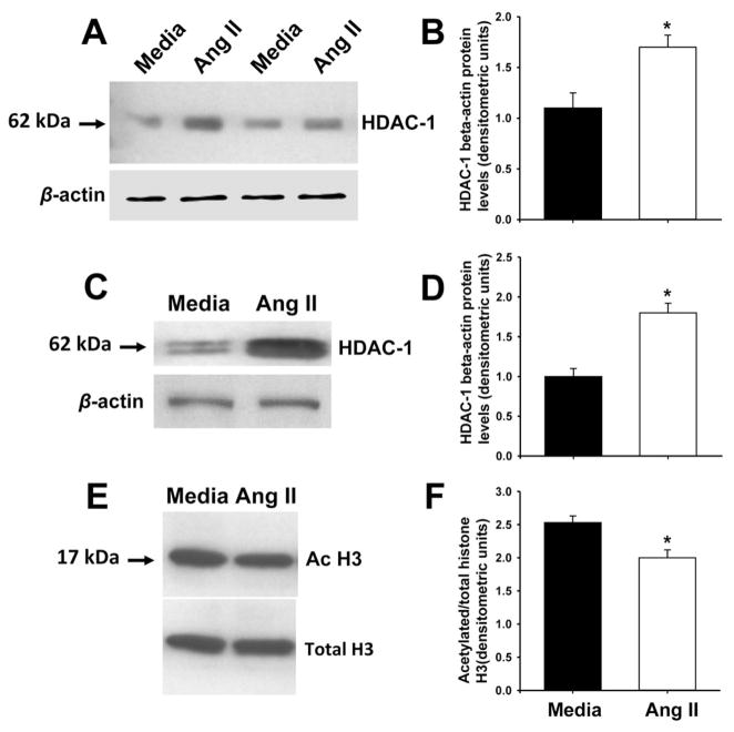
Mutations in the genes encoding components of the renin-angiotensin system (RAS) in mice or humans cause congenital abnormalities of the kidney and urinary tract. We hypothesized that absence of angiotensin (Ang) II in angiotensinogen (AGT)-deficient mice leads to defects in ureteric bud (UB) branching and that RAS genes are critically dependent on histone deacetylase (HDAC) activity. The number of UB tips was lower in AGT-/- compared with AGT+/+ embryonic (E) day E13.5 metanephroi (24+/-1.5 versus 36+/-3.7, p<0.05). Real-time RT-PCR demonstrated that pharmacological inhibition of HDAC activity with Scriptaid increases AGT, renin, angiotensin-converting enzyme (ACE), and AT1 receptor (AT1R) mRNA levels in E12.5 mouse metanephroi and early mesenchymal (MK3) cells. Furthermore, Scriptaid enhanced Ang II induced decrease in Sprouty (Spry) 1 gene expression in cultured UB cells. Treatment of intact E12.5 mouse metanephroi grown ex vivo with Ang II (10(-5) M, 24 h) increased HDAC-1 and decreased total acetylated histone H3 protein levels. These findings indicate that lack of endogenous Ang II in AGT-deficient mice inhibits UB branching. We conclude that intact RAS is critical in structural integrity of the renal collecting system and that UB morphogenetic program genes, such as AGT, renin, ACE, AT1R, or Spry1, are epigenetically controlled via HDACs.
Figures


Similar articles
-
Angiotensin II AT2 receptor regulates ureteric bud morphogenesis.Am J Physiol Renal Physiol. 2010 Mar;298(3):F807-17. doi: 10.1152/ajprenal.00147.2009. Epub 2009 Dec 23. Am J Physiol Renal Physiol. 2010. PMID: 20032120 Free PMC article.
-
Foxd1 is an upstream regulator of the renin-angiotensin system during metanephric kidney development.Pediatr Res. 2017 Nov;82(5):855-862. doi: 10.1038/pr.2017.157. Epub 2017 Aug 2. Pediatr Res. 2017. PMID: 28665931 Free PMC article.
-
A new role for the renin-angiotensin system in the development of the ureteric bud and renal collecting system.Keio J Med. 2008 Dec;57(4):184-9. doi: 10.2302/kjm.57.184. Keio J Med. 2008. PMID: 19110530 Free PMC article. Review.
-
Angiotensin II-induced activation of c-Ret signaling is critical in ureteric bud branching morphogenesis.Mech Dev. 2010 Jan-Feb;127(1-2):21-7. doi: 10.1016/j.mod.2009.11.004. Epub 2009 Dec 2. Mech Dev. 2010. PMID: 19961928 Free PMC article.
-
Renin-angiotensin system in ureteric bud branching morphogenesis: implications for kidney disease.Pediatr Nephrol. 2014 Apr;29(4):609-20. doi: 10.1007/s00467-013-2616-3. Epub 2013 Sep 7. Pediatr Nephrol. 2014. PMID: 24061643 Review.
Cited by
-
Vascular versus tubular renin: role in kidney development.Am J Physiol Regul Integr Comp Physiol. 2015 Sep 15;309(6):R650-7. doi: 10.1152/ajpregu.00313.2015. Epub 2015 Aug 5. Am J Physiol Regul Integr Comp Physiol. 2015. PMID: 26246508 Free PMC article.
-
(Pro)renin Receptor in Kidney Development and Disease.Int J Nephrol. 2011;2011:247048. doi: 10.4061/2011/247048. Epub 2011 Jun 20. Int J Nephrol. 2011. PMID: 21755055 Free PMC article.
-
Preterm Birth and Kidney Health: From the Womb to the Rest of Life.Children (Basel). 2024 Oct 2;11(10):1213. doi: 10.3390/children11101213. Children (Basel). 2024. PMID: 39457178 Free PMC article. Review.
-
Human GRK4γ142V Variant Promotes Angiotensin II Type I Receptor-Mediated Hypertension via Renal Histone Deacetylase Type 1 Inhibition.Hypertension. 2016 Feb;67(2):325-34. doi: 10.1161/HYPERTENSIONAHA.115.05962. Epub 2015 Dec 14. Hypertension. 2016. PMID: 26667412 Free PMC article.
-
Trichostatin A Modulates Angiotensin II-induced Vasoconstriction and Blood Pressure Via Inhibition of p66shc Activation.Korean J Physiol Pharmacol. 2015 Sep;19(5):467-72. doi: 10.4196/kjpp.2015.19.5.467. Epub 2015 Aug 20. Korean J Physiol Pharmacol. 2015. PMID: 26330760 Free PMC article.
References
-
- Saxen L. Organogenesis of the Kidney. Cambridge University Press; Cambridge: 1987.
-
- Ekblom P. Developmentally regulated conversion of mesenchyme to epithelium. FASEB J. 1989;3:2141–2150. - PubMed
-
- Grobstein C. Inductive epithelio-mesenchymal interaction in cultured organ rudiments of the mouse metanephros. Science. 1953;118:52–55. - PubMed
-
- Sakurai H, Nigam S. In vitro branching tubulogenesis: implications for developmental and cystic disorders, nephron number, renal repair, and nephron engineering. Kidney Int. 1998;54:14–26. - PubMed
-
- Brenner BM, Garcia DL, Anderson S. Glomeruli and blood pressure. Less of one, more the other? Am J Hypertens. 1988;1:335–347. - PubMed
Publication types
MeSH terms
Substances
Grants and funding
LinkOut - more resources
Full Text Sources
Research Materials
Miscellaneous
