

A case of gangliocytic paraganglioma in the ampulla of Vater
- PMID: 20497533
- PMCID: PMC2887868
- DOI: 10.1186/1477-7819-8-42
A case of gangliocytic paraganglioma in the ampulla of Vater
Abstract
Background: Duodenal gangliocytic paraganglioma is an extremely rare tumor and few cases have been reported to date.
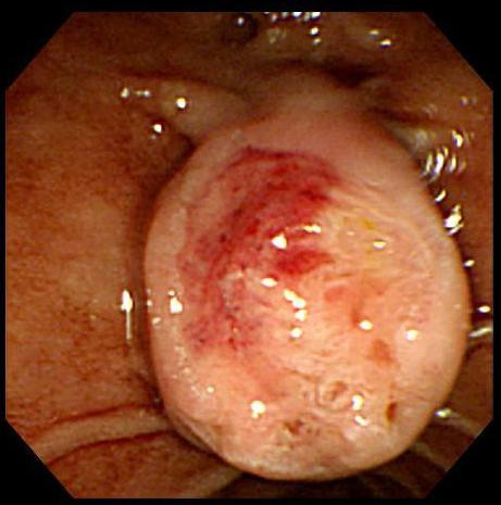
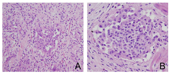
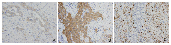
Case presentation: The authors report a case of gangliocytic paraganglioma verified by post-op pathology after pancreaticoduodenectomy for a tumor in the ampulla of Vater. The 56-year-old male patient concerned visited our emergency room with melena that started one week prior to hospitalization. The patient was diagnosed to have a tumor in the ampulla of Vater with bleeding on its surface. However post-op, he was diagnosed as having gangliocytic paraganglioma by immunohistochemistry.
Conclusion: This tumor has precise clinical implications, and if continuous follow up is conducted after careful diagnosis and surgical treatment, invasive major operations, such as, radical pancreaticoduodenectomy can be avoided.
Figures
References
-
- Yoo CY, Jung CK, Song KY, Kim SW, Lee KY. Gangliocytic Paraganglioma of the Duodenum. J Korean Surg Soc. 2007;73:68–71.
-
- Stanley RH, Lauri AA. WHO Classification of Tumors: Pathology and Genetics of Tumors of Digestive System. 1. Lyon: IARC press; 2000.
-
- Burke AP, Helwig EB. Gangliocytic paraganglioma. Am J Clin Pathol. 1989;92:1–9. - PubMed
Publication types
MeSH terms
Substances
LinkOut - more resources
Full Text Sources
Miscellaneous
