

Brain connectivity is not only lower but different in schizophrenia: a combined anatomical and functional approach
- PMID: 20497901
- PMCID: PMC2900394
- DOI: 10.1016/j.biopsych.2010.03.035
Brain connectivity is not only lower but different in schizophrenia: a combined anatomical and functional approach
Abstract
Background: Schizophrenia is hypothesized to involve disordered connectivity between brain regions. Currently, there are no direct measures of brain connectivity; functional and structural connectivity used separately provide only limited insight. Simultaneous measure of anatomical and functional connectivity and its interactions allow for better understanding of schizophrenia-related alternations in brain connectivity.
Methods: Twenty-seven schizophrenia patients and 27 healthy control subjects underwent magnetic resonance imaging with resting state functional magnetic resonance imaging and diffusion tensor imaging. Separate functional and anatomical connectivity maps were calculated and combined for each subject. Global, regional, and voxel measures and K-means network analysis were employed to identify group differences and correlation with clinical symptoms.
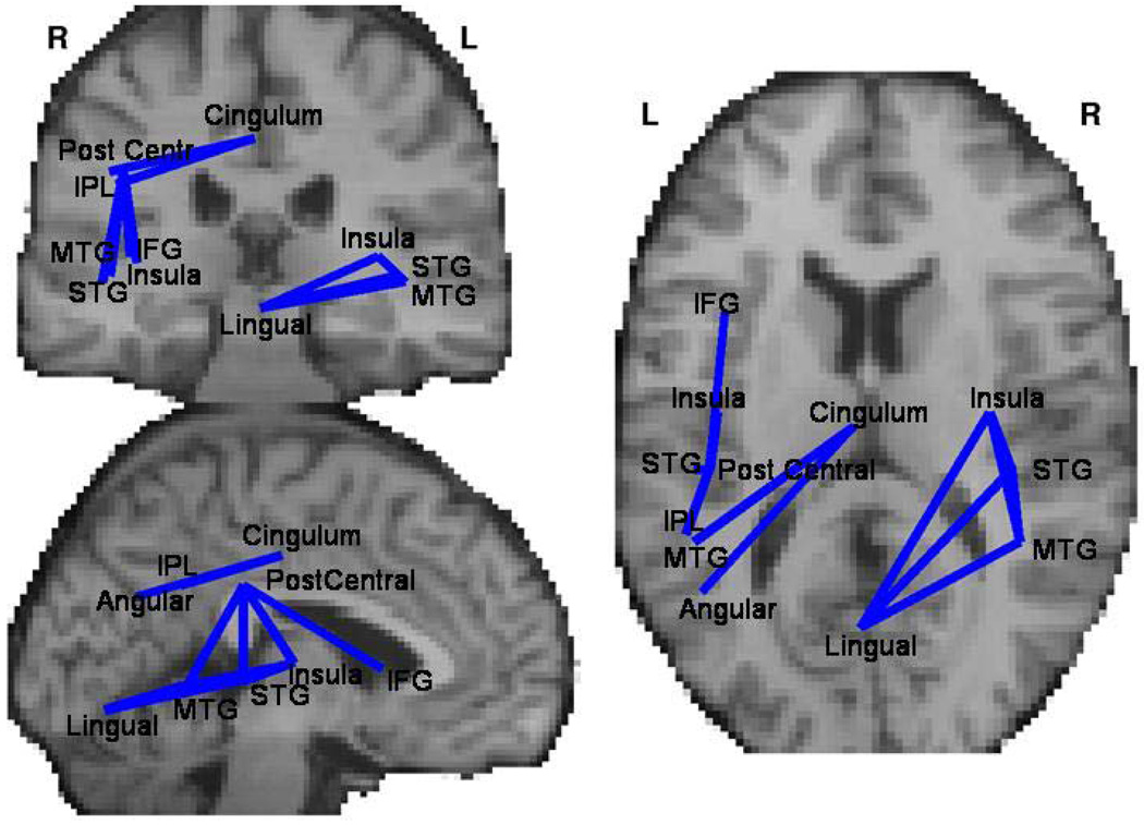
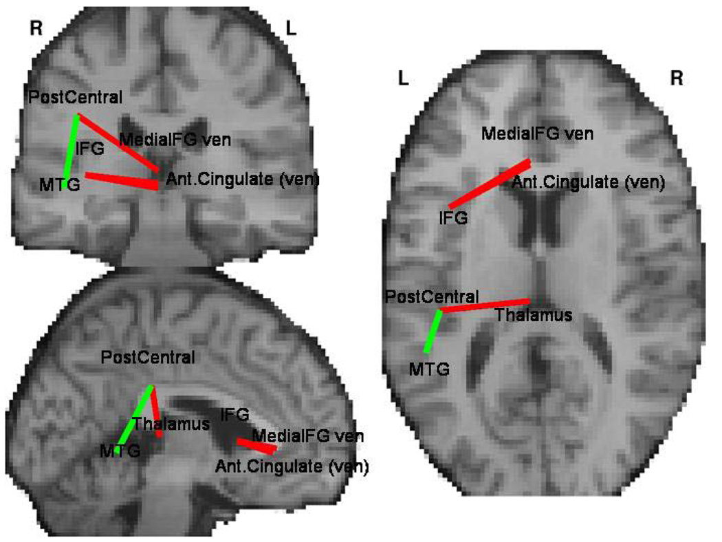
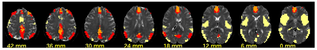
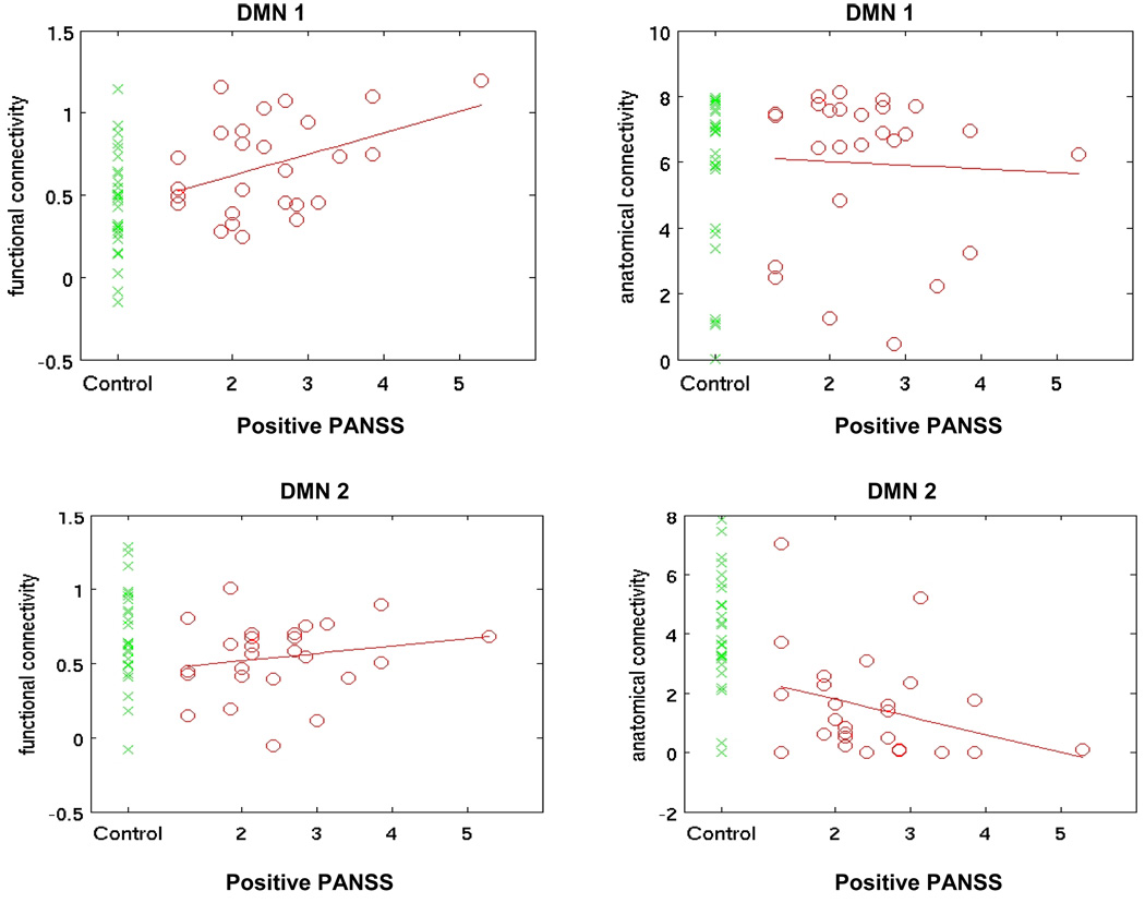
Results: A global connectivity analysis indicated that patients had lower anatomical connectivity and lower coherence between the two imaging modalities. In schizophrenia these group differences correlated with clinical symptom severity. Although anatomical connectivity nearly uniformly decreased, functional connectivity in schizophrenia was lower for some connections (e.g., middle temporal gyrus) and higher for others (e.g., cingulate and thalamus). Within the default mode network (DMN) two separate subsystems can be identified. Schizophrenia patients showed decoupling between structural and functional connectivity that can be localized to networks originating in posterior cingulate cortex as well as in the task-positive network and one of the DMN components.
Conclusions: Combining two measures of brain connectivity provides more comprehensive descriptions of altered brain connectivity underlying schizophrenia. Patients show deficits in white matter anatomy, but functional connectivity alterations are more complex. Fusion of both methods allows identification of subsystems showing both increased and decreased functional connectivity.
Copyright 2010 Society of Biological Psychiatry. Published by Elsevier Inc. All rights reserved.
Figures
References
-
- Bleuler E. Dementia Praecox or the Group of Schizophrenias 9. International Universities Press; 1911.
-
- Volkow ND, Brodie JD, Wolf AP, Gomezmont F, Cancro R, Vangelder P, et al. BRAIN ORGANIZATION IN SCHIZOPHRENIA. Journal of Cerebral Blood Flow and Metabolism. 1986;6:441–446. - PubMed
-
- Friston KJ. Schizophrenia and the disconnection hypothesis. Acta Psychiatrica Scandinavica. 1999;99:68–79. - PubMed
Publication types
MeSH terms
Grants and funding
LinkOut - more resources
Full Text Sources
Other Literature Sources
Medical
