. 2010 Aug;116(2):623-31.
doi: 10.1093/toxsci/kfq157. Epub 2010 May 23.

Development of a prolonged calcium plateau in hippocampal neurons in rats surviving status epilepticus induced by the organophosphate diisopropylfluorophosphate

Affiliations

Development of a prolonged calcium plateau in hippocampal neurons in rats surviving status epilepticus induced by the organophosphate diisopropylfluorophosphate

Laxmikant S Deshpande et al. Toxicol Sci. 2010 Aug.

Abstract

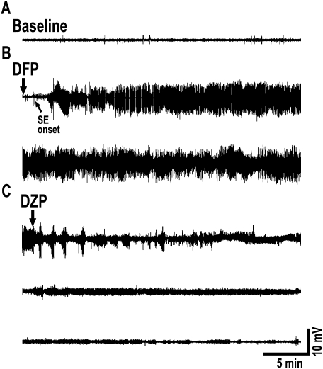
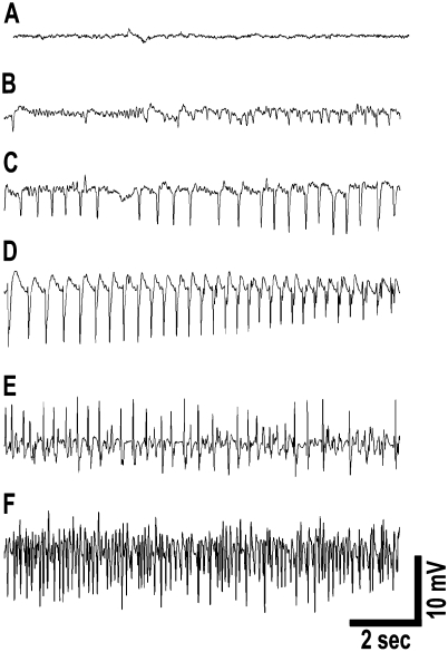
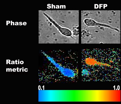
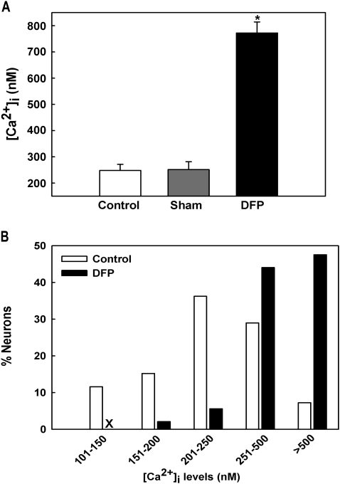
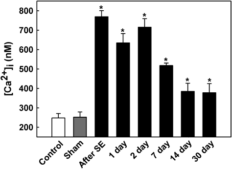
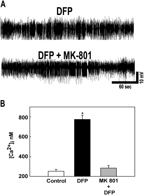
Organophosphate (OP) compounds are among the most lethal chemical weapons ever developed and are irreversible inhibitors of acetylcholinesterase. Exposure to majority of OP produces status epilepticus (SE) and severe cholinergic symptoms that if left untreated are fatal. Survivors of OP intoxication often suffer from irreversible brain damage and chronic neurological disorders. Although pilocarpine has been used to model SE following OP exposure, there is a need to establish a SE model that uses an OP compound in order to realistically mimic both acute and long-term effects of nerve agent intoxication. Here we describe the development of a rat model of OP-induced SE using diisopropylfluorophosphate (DFP). The mortality, behavioral manifestations, and electroencephalogram (EEG) profile for DFP-induced SE (4 mg/kg, sc) were identical to those reported for nerve agents. However, significantly higher survival rates were achieved with an improved dose regimen of DFP and treatment with pralidoxime chloride (25 mg/kg, im), atropine (2 mg/kg, ip), and diazepam (5 mg/kg, ip) making this model ideal to study chronic effects of OP exposure. Further, DFP treatment produced N-methyl-D-aspartate (NMDA) receptor-mediated significant elevation in hippocampal neuronal [Ca(2+)](i) that lasted for weeks after the initial SE. These results provided direct evidence that DFP-induced SE altered Ca(2+) dynamics that could underlie some of the long-term plasticity changes associated with OP toxicity. This model is ideally suited to test effective countermeasures for OP exposure and study molecular mechanisms underlying neurological disorders following OP intoxication.


Figures

FIG. 1.
FIG. 1.
EEG seizure activity evoked by DFP. (A) Baseline activity before administration of DFP. (B) Onset of DFP action is rapid producing rhythmic high-amplitude and high-frequency spike activity that rapidly progresses into electrographic SE. SE is robust, intense, and does not wax and wane for the entire 1 h. The two traces in panel B together represents one full hour of electrographic SE. (C) Seizures are terminated with diazepam (DZP) injections at 1, 3, and 5 h following onset of SE. These treatments completely stop EEG epileptiform activity. The three traces in panel C represent EEG activity following the three diazepam injections. Traces are representative of n = 6 animals.
FIG. 2.
FIG. 2.
EEG progression of DFP-induced SE. (A) Baseline activity prior to DFP injection. (B) Low-voltage fast activity appears within 2–3 min of DFP administration. (C) High-voltage slow activity appears within the next minute that progresses to (D) high-frequency and high-voltage spiking and (E and F) sustained continuous seizure activity. EEG activity at 15 min following DFP administration (F) demonstrates electrographic SE with fully developed continuous seizures. Spike frequency during SE was sustained at greater than 7–10 Hz. SE was induced by DFP within 7–8 min following injections. EEG activity correlated with behavioral observations.
FIG. 3.
FIG. 3.
Phase contrast and pseudocolor ratiometric images of representative sham- and DFP-treated neurons. Sham-treated neurons had bluish color that corresponds to lower Fura-2 ratio whereas DFP-treated neurons had orange-red color that corresponds to high Fura-2 ratio.
FIG. 4.
FIG. 4.
Basal [Ca2+]i in acutely isolated CA1 hippocampal neurons following DFP-induced SE. (A) Elevated [Ca2+]i in CA1 hippocampal neurons acutely isolated from animals that had experienced 1 h of DFP-induced SE compared with neurons from control and sham-treated animals. (*p < 0.001, one-way ANOVA, post hoc Tukey test, n = 7, 5, and 6 animals, respectively). Data are represented as mean ± SEM. (B) Distribution of [Ca2+]i levels for sham and DFP-SE hippocampal neurons. Sham neurons demonstrated a normal distribution for [Ca2+]i levels with approximately 90% of neurons exhibiting [Ca2+]i levels less than 500nM and only 7–10% neurons exhibiting very high [Ca2+]i levels. In contrast, DFP-SE neurons demonstrated a rightward shift toward higher [Ca2+]i levels with approximately 50% neurons exhibiting [Ca2+]i levels greater than 500nM (n = 150 neurons for each condition).
FIG. 5.
FIG. 5.
Development of Ca2+ plateau following DFP-induced SE. CA1 hippocampal [Ca2+]i in control (white bar), sham (gray bar), and DFP immediately after and 1, 2, 7, 14, and 30 days after SE (black bars). [Ca2+]i in sham animals at each time point were not significantly different from the control values shown and thus were omitted from the graph for clarity. [Ca2+]i in neurons isolated from DFP-SE animals was significantly higher than control and sham values (*p < 0.05, one-way ANOVA, post hoc Tukey test, n = minimum of six animals at each time). Data are represented as mean ± SEM.
FIG. 6.
FIG. 6.
Effect of MK-801 administration on DFP-induced SE. (A) Representative EEG pattern from animals pretreated with MK-801 and DFP alone. MK-801 pretreatment did not affect the severity, intensity, and duration of DFP-induced SE. (B) MK-801 pretreatment prevented the elevations in [Ca2+]i that occur following DFP-induced SE. (*p < 0.01, one-way ANOVA, post hoc Tukey test, n = 5, 7, and 6 animals, respectively). Data are represented as mean ± SEM.

References

    1. Acharya J, Gupta AK, Dubey DK, Raza SK. Synthesis and evaluation of novel bis-pyridinium oximes as reactivators of DFP-inhibited acetylcholinesterase. Eur. J. Med. Chem. 2009;44:1335–1340. - PubMed
    1. Antonijevic B, Stojiljkovic MP. Unequal efficacy of pyridinium oximes in acute organophosphate poisoning. Clin. Med. Res. 2007;5:71–82. - PMC - PubMed
    1. Auta J, Costa E, Davis J, Guidotti A. Imidazenil: a potent and safe protective agent against diisopropyl fluorophosphate toxicity. Neuropharmacology. 2004;46:397–403. - PubMed
    1. Bajgar J. Organophosphates/nerve agent poisoning: mechanism of action, diagnosis, prophylaxis, and treatment. Adv. Clin. Chem. 2004;38:151–216. - PubMed
    1. Bouzarth WF, Himwich HE. Mechanism of seizures induced by di-isopropyl flurophosphate (DFP) Am. J. Psychiatry. 1952;108:847–855. - PubMed

Publication types