

doi: 10.1073/pnas.1000525107.
Epub 2010 May 24.
The Mohawk homeobox gene is a critical regulator of tendon differentiation
Affiliations
- PMID: 20498044
- PMCID: PMC2890854
- DOI: 10.1073/pnas.1000525107
Item in Clipboard
The Mohawk homeobox gene is a critical regulator of tendon differentiation
Proc Natl Acad Sci U S A.
.
Abstract
Mohawk (Mkx) is a member of the Three Amino acid Loop Extension superclass of atypical homeobox genes that is expressed in developing tendons. To investigate the in vivo functions of Mkx, we generated Mkx(-/-) mice. These mice had hypoplastic tendons throughout the body. Despite the reduction in tendon mass, the cell number in tail tendon fiber bundles was similar between wild-type and Mkx(-/-) mice. We also observed small collagen fibril diameters and a down-regulation of type I collagen in Mkx(-/-) tendons. These data indicate that Mkx plays a critical role in tendon differentiation by regulating type I collagen production in tendon cells.
Conflict of interest statement
The authors declare no conflict of interest.
Figures
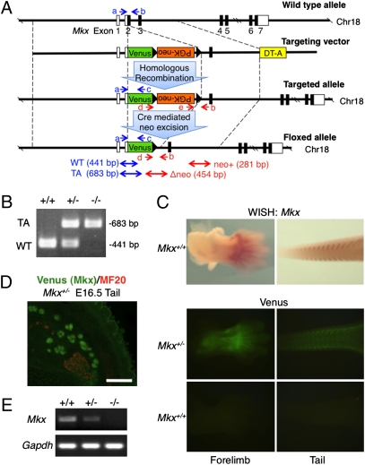
Generation of Mkx mutant mice. (A) Diagram of the Mkx targeting construct. Blue and red arrows (a–e) show genomic PCR primers for genotyping. White box, UTR; Black box, coding region; DT-A, diphtheria toxin A; WT, wild-type allele; TA, targeted allele. (B) Genomic PCR of wild-type and Mkx mutant mice for genotyping using primers a, b and c. (C) Whole-mount in situ hybridization of Mkx (Upper) and whole-mount visualization of Venus signals (Lower) in E13.5 forelimb and tail of wild-type or Mkx mutant embryos. (D) Immunohistochemistry for anti-myosin heavy chain (MF20; red) and visualization of Venus in cryosection of E16.5 tail of a Venus knockin Mkx heterozygous embryo. (Scale bar, 100 μm.) (E) RT-PCR analysis for Mkx and Gapdh of Achilles tendon in wild-type and Mkx mutant mice.
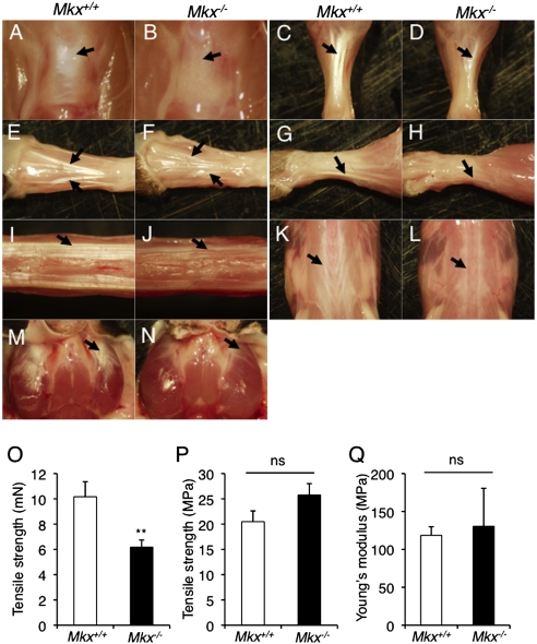
Tendon defects are observed in Mkx null mice. (A–N) The appearance of the patellar tendon (A and B; black arrow), Achilles tendon (C and D; black arrow), hindlimb tendons (E and F), forelimb tendons (G and H), tail tendons (I and J), back tendons (K and L; black arrow) and platysma tendon (M and N; black arrow) in 3-month-old wild-type and Mkx null mice. (O and P) Absolute value of tensile strength (O) and tensile strength per unit area (P) of Achilles tendons in wild-type and Mkx null mice. Error bars, SEM (n = 7). ns, no significance. (Q) Young's modulus of Achilles tendons in wild-type and Mkx null mice. Error bars, SEM (n = 7). ns, no significance.
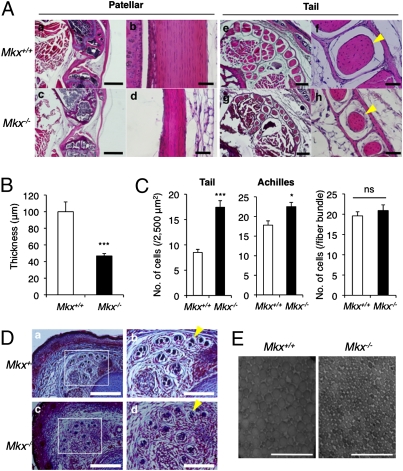
Tendon mass is decreased in Mkx null mice. (A) H&E staining of the patellar (a–d) and tail (e–h) tendons in 3-month-old wild-type and Mkx null mice. Yellow arrowheads indicate a fiber bundle in the tail tendon. [Scale bars: (a and c) 1 mm; (b and d) 50 μm; (e and g) 200 μm; (f and h) 50 μm.] (B) Patellar tendon thickness of 7-day-old wild-type and Mkx null mice. Error bars, SEM (n = 8). (C) Cell density of tail or Achilles tendons (Left) and cell number in a tail tendon fiber bundle (Right) in 3-month-old wild-type and Mkx null mice. Error bars, SEM (Left, n = 8 ~10; Right, n = 14). ns, no significance. (D) Azan staining of E18.5 embryonic tails in wild-type and Mkx null mice. Yellow arrowheads indicate a fiber bundle in the tail tendon. [Scale bars: (Left) 500 μm; (Right) 200 μm.] (E) Transmission electron microscopic view of collagen fibrils in the Achilles tendon of wild-type and Mkx null mice. (Scale bars, 200 nm.)
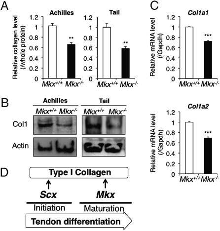
Type I collagen productivity is decreased in Mkx null tendon cells. (A) Soluble collagen measurement in whole soluble protein of Achilles or tail tendons of 8-week-old wild-type or Mkx null mice. Error bars, SEM (n = 3). (B) Western blot analysis of type I collagen (Col1) and β-actin (actin) in Achilles or tail tendons of 8-week-old wild-type or Mkx null mice. (C) Real-time PCR analysis for Col1a1 and Col1a2 in Achilles tendons of 8-week-old wild-type or Mkx null mice. Error bars, SEM (n = 3). (D) Proposed tendon differentiation network.
References
-
- Sharma P, Maffulli N. Biology of tendon injury: Healing, modeling and remodeling. J Musculoskelet Neuronal Interact. 2006;6:181–190. - PubMed
-
- Bi Y, et al. Identification of tendon stem/progenitor cells and the role of the extracellular matrix in their niche. Nat Med. 2007;13:1219–1227. - PubMed
-
- Kannus P. Structure of the tendon connective tissue. Scand J Med Sci Sports. 2000;10:312–320. - PubMed
-
- Yoon JH, Halper J. Tendon proteoglycans: Biochemistry and function. J Musculoskelet Neuronal Interact. 2005;5:22–34. - PubMed
Publication types
MeSH terms
Substances
LinkOut - more resources
Full Text Sources
Other Literature Sources
Molecular Biology Databases
Research Materials
