. 2010 Jun 8;107(23):10490-5.
doi: 10.1073/pnas.1002128107. Epub 2010 May 24.

Tubulin polyglutamylation is essential for airway ciliary function through the regulation of beating asymmetry

Affiliations

Tubulin polyglutamylation is essential for airway ciliary function through the regulation of beating asymmetry

Koji Ikegami et al. Proc Natl Acad Sci U S A. .

Abstract

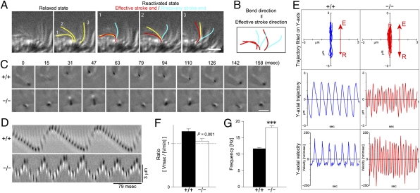
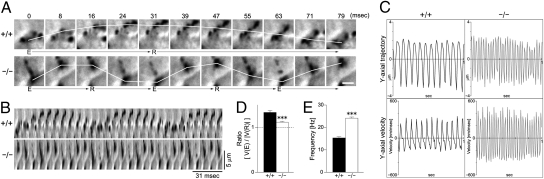
Airway epithelial cilia protect the mammalian respiratory system from harmful inhaled materials by providing the force necessary for effective mucociliary clearance. Ciliary beating is asymmetric, composed of clearly distinguished effective and recovery strokes. Neither the importance of nor the essential components responsible for the beating asymmetry has been directly elucidated. We report here that the beating asymmetry is crucial for ciliary function and requires tubulin glutamylation, a unique posttranslational modification that is highly abundant in cilia. WT murine tracheal cilia have an axoneme-intrinsic structural curvature that points in the direction of effective strokes. The axonemal curvature was lost in tracheal cilia from mice with knockout of a tubulin glutamylation-performing enzyme, tubulin tyrosine ligase-like protein 1. Along with the loss of axonemal curvature, the axonemes and tracheal epithelial cilia from these knockout (KO) mice lost beating asymmetry. The loss of beating asymmetry resulted in a reduction of cilia-generated fluid flow in trachea from the KO mice. The KO mice displayed a significant accumulation of mucus in the nasal cavity, and also emitted frequent coughing- or sneezing-like noises. Thus, the beating asymmetry is important for airway ciliary function. Our findings provide evidence that tubulin glutamylation is essential for ciliary function through the regulation of beating asymmetry, and provides insight into the molecular basis underlying the beating asymmetry.


Conflict of interest statement

The authors declare no conflict of interest.

Figures

Fig. 1.
Fig. 1.
Generation of Ttll1-KO mice. (A and B) Generation of Ttll1-deficient mice. (A) Exons 2–4 were replaced by a TKneo cassette through homologous recombination. (B) Each mouse was genotyped by PCR with primers depicted in A as white arrows. (C) Western blot analysis of TTLL1. TTLL1 protein was absent in the trachea and lung of Ttll1-deficent homozygote (−/−) mice. (D) Results of heterozygous mating. The graph shows the ratio of each genotype [WT (+/+), heterozygote (+/−), or KO homozygote (−/−)] born from heterozygous mating. In total, 690 animals obtained from 95 matings were genotyped. P = 0.279, χ2 test. (E) Immunostaining of tracheal tissue section. WT cilia were stained by a mAb against glutamylated tubulin (GT335, green), whereas cilia from Ttll1-KO mice were not. (Scale bar: 5 μm.) (F) Detection of glutamylated tubulins in trachea. Ttll1-KO mice showed greatly reduced levels of glutamylated (PG) tubulin detected by the mAb GT335. Differences of other modifications—acetylation (Ac), tyrosination (Tyr), detyrosination (Detyr), and Δ2—were subtle when present. (G) Immunostaining of isolated ciliary axonemes with the mAb GT335. (Scale bar: 5 μm.) Arrowheads point to straight axonemes. (H) Detection of glutamylated tubulins and other modifications in isolated ciliary axonemes.
Fig. 2.
Fig. 2.
Loss of structural curvature in ciliary axonemes of Ttll1-KO mice. (A) Phase-contrast micrograph of isolated ciliary axonemes. Arrows point to straightened axonemes in the Ttll1-KO preparation. (Scale bar: 5 μm.) (B and C) Bend angles of isolated ciliary axonemes. Both the histogram (B) and the boxplot (C) of bend angles show that the majority of Ttll1-KO axonemes had bend angles <40 degrees. In C, data are presented as median ± quartile (box) and 90th percentile (bars). ***P < 0.001, Mann-Whitney U test [n = 621 (+/+), 557 (−/−)]. (D and E) Bend angles of relaxed axonemes. Both the histogram (D) and the boxplot (E) of bend angles show that the majority of Ttll1-KO axonemes had bend angles <40 degrees. In E, data are presented as median ± quartile (box) and 90th percentile (bars). ***P < 0.001, Mann-Whitney U test [n = 244 (+/+), 186 (−/−)]. (F) Boxplot of lengths of ciliary axonemes. Data are presented as median ± quartile (box) and 90th percentile (bars). No statistically significant difference was detected. (G) Representative shape of axonemes depicted from medians of angle and length. The overlay image clearly shows the reduced bend angle of the Ttll1-KO axonemes.
Fig. 3.
Fig. 3.
Loss of beating asymmetry in Ttll1-KO ciliary axonemes. (A and B) Relationship between bend direction and beating direction. (A) Three representative cilia are highlighted. In the relaxed state, all cilia retained bends to the left (yellow). After reactivation, the cilia beat to the left (blue to red). (Scale bar: 5 μm.) (B) Note that the bend direction is the same as the direction of the effective stroke. (C) Time-lapse photography of ATP-reactivated ciliary axoneme. (Scale bar: 3 μm.) (D) Kymograph of an ATP-reactivated ciliary axoneme. (E) Analyses of axonemal motility. The beating plane of the original trajectory was fitted on the y axis (Top). The y-axial trajectory was plotted as a time function (Middle). The velocity in the y-direction was plotted against time (Bottom). (F) Distinction between effective and recovery strokes. The ratio between the maximum and minimum velocity was decreased significantly in axonemes from the Ttll1-KO mice and approached a value of 1, indicating a symmetric beating pattern [P = 0.001, one-way ANOVA; n = 56 (+/+), 65 (−/−)]. (G) Frequency of axonemal beating. Axonemes isolated from Ttll1-KO mice showed a significantly increased ciliary beating frequency compared with axonemes isolated from WT animals (P < 0.001, one-way ANOVA).
Fig. 4.
Fig. 4.
Loss of beating asymmetry of tracheal epithelial cilia in Ttll1-KO mice. (A) Time-lapse photography of labeled cilia. The white line shows a trace of ciliary motility during the effective (E) and recovery (R) strokes. (Scale bar: 3 μm.) (B) Kymograph of ciliary strokes. (C) Analyses of ciliary motility. The ciliary trajectory was plotted as a time function after being fitted onto the y axis (Upper). The velocity in the y-direction was plotted against time (Lower). (D) Distinction between effective and recovery strokes. The ratio of velocities between effective and recovery strokes was significantly decreased in Ttll1-KO cilia (P < 0.001, one-way ANOVA; n = 40). (E) Frequency of ciliary beating. Ttll1-KO cilia showed a significantly higher beating frequency than WT cilia (P < 0.001, one-way ANOVA).
Fig. 5.
Fig. 5.
Ciliary dysfunction, rhinosinusitis, and respiratory defects in Ttll1-KO mice. (A and B) Cilia-generated flow. In A, 500-ms trajectories of polystyrene beads deposited on the tracheal surface are depicted in 66-ms intervals. (Scale bar: 10 μm.) Quantified data are shown in B. The velocity of each bead was estimated by dividing the width of the field of view (50 μm) by the time each individual bead takes to pass through the field. Data are presented as mean ± SEM [n = 15 (+/+), 10 (−/−)]. ***P < 0.001, one-way ANOVA. (C) Orientation of ciliary beating. The mean deviation showed a slight, but statistically significant increase in Ttll1-KO mice. P values were analyzed with the Mann-Whitney U test [n = 279 (+/+), 304 (−/−)]. (D and E) Orientation of ciliary basal feet. (D) The basal feet of both WT and Ttll1-KO mice point in the same direction (white arrows) in the transmission electron microscopy (TEM) micrographs. (E) Rose diagrams of basal foot orientation. There was no statistically significant difference using the Mann-Whitney U test [n = 121 (+/+), 146 (−/−)]. MD, mean deviation. (F) Alcian blue (Top) and H&E (Middle) staining of nasal cavity. In Ttll1-KO mice, accumulated mucus was clearly visible (asterisks), whereas WT mice showed no mucus accumulation. (Bottom) A magnified photograph of the boxed region of the H&E-stained KO sample. (Inset) Neutrophils in the nasal cavity of a Ttll1-KO mouse. [Scale bars: 0.4 mm (Top and Middle), 50 μm (Bottom).] (G and H) Coughing-/sneezing-like phenotypes in Ttll1-KO mice. (G) Strong spikes (arrows) were observed only in spectrograms recorded from KO animals. (H) Quantification of the frequency of coughing-/sneezing-like noises observed in Ttll1-KO mice. ***P < 0.001, one-way ANOVA (n = 3). No coughing-/sneezing-like noises were recorded from WT mice.

Similar articles

Cited by

References

    1. Gibbons IR. The relationship between the fine structure and direction of beat in gill cilia of a lamellibranch mollusc. J Biophys Biochem Cytol. 1961;11:179–205. - PMC - PubMed
    1. Afzelius BA. A human syndrome caused by immotile cilia. Science. 1976;193:317–319. - PubMed
    1. Ferkol T, Leigh M. Primary ciliary dyskinesia and newborn respiratory distress. Semin Perinatol. 2006;30:335–340. - PubMed
    1. Morillas HN, Zariwala M, Knowles MR. Genetic causes of bronchiectasis: Primary ciliary dyskinesia. Respiration. 2007;74:252–263. - PubMed
    1. Nicastro D, et al. The molecular architecture of axonemes revealed by cryoelectron tomography. Science. 2006;313:944–948. - PubMed

Publication types