. 2010 Jun 8;107(23):10520-5.
doi: 10.1073/pnas.1001896107. Epub 2010 May 24.

Vascular Robo4 restricts proangiogenic VEGF signaling in breast

Affiliations

Vascular Robo4 restricts proangiogenic VEGF signaling in breast

Rebecca Marlow et al. Proc Natl Acad Sci U S A. .

Abstract

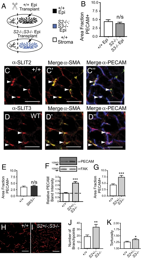
Formation of the vascular system within organs requires the balanced action of numerous positive and negative factors secreted by stromal and epithelial cells. Here, we used a genetic approach to determine the role of SLITs in regulating the growth and organization of blood vessels in the mammary gland. We demonstrate that vascularization of the gland is not affected by loss of Slit expression in the epithelial compartment. Instead, we identify a stromal source of SLIT, mural cells encircling blood vessels, and show that loss of Slit in the stroma leads to elevated blood vessel density and complexity. We examine candidate SLIT receptors, Robo1 and Robo4, and find that increased vessel angiogenesis is phenocopied by loss of endothelial-specific Robo4, as long as it is combined with the presence of an angiogenic stimulus such as preneoplasia or pregnancy. In contrast, loss of Robo1 does not affect blood vessel growth. The enhanced growth of blood vessels in Robo4(-/-) endothelium is due to activation of vascular endothelial growth factor (VEGF)-R2 signaling through the Src and FAK kinases. Thus, our studies present a genetic dissection of SLIT/ROBO signaling during organ development. We identify a stromal, rather than epithelial, source of SLITs that inhibits blood vessel growth by signaling through endothelial ROBO4 to down-regulate VEGF/VEGFR2 signaling.


Conflict of interest statement

Conflict of interest statement: D.Y.L. is employed by the University of Utah, which has filed intellectual property surrounding the therapeutic uses of targeting Robo4 and with the intent to license this body of intellectual property for commercialization.

Figures

Fig. 1.
Fig. 1.
Loss of global, but not epithelial Slits, enhances blood vessel growth. (A) Diagram illustrating transplants that generate chimeric mammary glands with Slit2−/−;Slit3−/− epithelium (blue) and contralateral WT epithelium (black), transplanted into immunocompromised (Foxn1nu) hosts (white) that have been precleared of their WT epithelium (black). (B) Lack of Slit in the epithelium does not alter blood vessel density in outgrowths. Quantitative analysis of PECAM-positive pixel area (n = 3 contralateral outgrowths, 15 fields of view (FOV)/outgrowth). Error bars = SEM. n/s = not significant. (C and D) Mural cells express SLIT2 and SLIT3. Representative images of sections immunostained for PECAM (blue), SMA (green), and SLIT2 or SLIT3 (red). Arrows indicate mural cell localization. (Scale bar, 50 μm.) (E) Lack of Slit3 does not increase blood vessel number in the mammary gland. Quantitative analysis of PECAM-positive pixel area (n = 3 animals, 15 FOV/gland). Error bars = SEM. n/s = not significant. (FK) Global lack of Slit significantly increases blood vessel number and network complexity. (F) Representative PECAM immunoblots on WT and Slit2+/−;Slit3−/− mammary lysates (50 μg loaded; FAK immunoblot is loading control). Bar graph represent quantitative analysis of PECAM band intensity (ImageJ) (n = 3). Error bars = SEM, ***P < 0.001 unpaired t test. (G) Quantitative analysis of PECAM-positive pixel area (n = 3 animals, 15 FOV/animal). Error bars = SEM. *** P < 0.001 unpaired t test. (H and I) Representative images of WT (H) and Slit2+/−;Slit3−/− (I) mammary sections immunostained with anti-PECAM (red). (Scale bar = 50 μm.) (J) Number of branchpoints and (K) tortuosity of blood vessels were quantified. Error bars = SEM, ***P < 0.001, **P < 0.005, *P < 0.01 unpaired t test.
Fig. 2.
Fig. 2.
Loss of ROBO1 and ROBO4 enhances blood vessel growth. (A) Lack of Robo4 does not alter blood vessel density in the mammary gland. Quantitative analysis of PECAM-positive pixel area (n = 3 animals, 15 FOV/animal). Error bars = SEM. n/s = not significant. (B) ROBO1 is expressed by blood vessels. Representative images of ROBO1 (green) and PECAM (red) immunostaining on WT mammary sections. (Scale bar, 20 μm.) (C) Lack of Robo1 does not alter blood vessel density in the mammary gland. Quantitative analysis of PECAM-positive pixel area (n = 3 animals, 15 FOV/animal). Error bars = SEM. n/s = not significant. (DI) Lack of both Robo1 and Robo4 significantly increases blood vessel density and network complexity in the mammary gland. (D) Representative PECAM immunoblots on WT, Robo1−/−, Robo4−/−, and Robo1−/−;Robo4−/− mammary lysates (50 μg loaded; FAK immunoblot is loading control). Bar graph represent quantitative analysis of PECAM band intensity (ImageJ) (n = 3). Error bars = SEM, ***P < 0.001 ANOVA. (E) Quantitative analysis of PECAM-positive pixel area (n = 4 animals, 15 FOV/animal). ***P < 0.001 unpaired t test. (F and G) Representative images of WT (F) and Robo1−/−;Robo4−/− (G) mammary sections immunostained with anti-PECAM (red). (Scale bar, 50 μm.) (H and I) Quantification of branchpoint number (H) and tortuosity (I). Error bars = SEM. ***P < 0.001, **P < 0.005 unpaired t test.
Fig. 3.
Fig. 3.
The proangiogenic factor, VEGF, emanating from Robo1−/− and pregnant epithelium increases angiogenesis in Robo4−/− glands. (A and B) SDF1 (A) and VEGF-A (B) are expressed at higher levels by Robo1−/− and Robo1−/−;Robo4−/−, compared to WT or Robo4−/−, mammary glands. Immunostained sections were scored according to cell percent positivity and staining intensity (n = 3 animals, 10 FOV/animal). Scores were plotted on a vertical scatter plot and red bars indicate average score. ***P < 0.001 ANOVA. (C) Diagram illustrating generation of chimeric glands with Robo1−/− epithelium (red) and contralateral WT epithelium (black), transplanted into immunocompromised (Foxn1nu) hosts (white) that have been cleared of their WT epithelium (black) (D) Lack of Robo1 in the epithelium does not alter blood vessel density in outgrowths. Quantification of PECAM-positive pixel area [n = 7 contralateral outgrowths, 15 FOV/outgrowth]. Error bars = SEM. n/s = not significant. (E) Diagram illustrating generation of chimeric glands with Robo1−/− epithelium (red) and contralateral WT epithelium (black) into syngeneic Robo4−/− background (light gray) that have been cleared of their host Robo4−/− epithelium (dark gray). (FH) Increased blood vessel density with loss of Robo1 in the epithelium combined with the loss of Robo4. (F) Quantitative analysis of PECAM-positive pixel area (n = 3 contralateral outgrowths, 15 FOV/outgrowth). Quantification of branchpoint number (G) and tortuosity (H). Error bars = SEM, **P < 0.005, *P < 0.01 unpaired t test. (I) Robo4 is necessary for SLIT2N-mediated inhibition of VEGF-induced human microvascular endothelial cells-lung (HMVEC-L) migration. HMVEC-L cells were subjected to control, Robo1, or Robo4 siRNA and allowed to migrate in response to VEGF in the presence of Mock or SLIT2N (n > 3, *** P < 0.001 ANOVA). Western blot analysis confirmed knockdown of Robo1 or Robo4 expression. (JM) Pregnancy increases blood vessel density in Robo4-/ glands compared to WT. (J) Representative immunoblots of anti-PECAM on WT and Robo4−/−mammary lysates (50 μg loaded; FAK immunoblot is loading control). Bar graph represents quantification of PECAM band intensity (ImageJ) (n = 3). Error bars = SEM, ***P < 0.001 unpaired t test. (K) Quantitative analysis of PECAM-positive pixel area (n = 3 animals, 10 FOV/animal). Error bars = SEM, **P < 0.005 unpaired t test. (L and M) Representative images of WT (L) and Robo4−/− (M) mammary sections from animals at pregnancy day 12.5 immunostained with anti-PECAM (red) and anti-CK14 (green), a marker for myoepithelial cells. Arrowheads indicate capillary baskets surrounding alveoli. (Scale bar, 50 μm.)
Fig. 4.
Fig. 4.
ROBO4 functions to restrain VEGF/VEGFR2 signaling in the mammary gland. (A) Increased activation of VEGFR2 in Robo1−/−;Robo4−/− but not WT, Robo1−/− or Robo4−/− glands. Immunoblotting for VEGFR2 and phosphotyrosine (4G10) after immunoprecipitation of VEGFR2 from adult gland lysates. Bar graph represents quantification of phospho-VEGFR2 relative to total VEGFR2 (ImageJ) (n = 4 per stage and per genotype). Error bars = SEM, **P < 0.005 unpaired t test. (B) Increased activation of VEGFR2 in Robo4−/− in pregnant glands (day 12.5) compared to WT. Bar graph represents quantification of phospho-VEGFR2 relative to total VEGFR2 (ImageJ) (n = 3). Error bars = SEM, **P < 0.005 unpaired t test. (C) Increased activation of VEGFR2 in Robo1−/−;Robo4−/− adult glands and Robo4−/− pregnant glands, compared to WT controls. Bar graphs represent area fraction of pixels positive for PY1175-VEGFR2 divided by the area fraction positive for PECAM (n = 4 animals/genotype, 10 FOV/animal). Error bars = SEM, **P < 0.005 ANOVA. (D) Increased activation of Src in Robo1−/−;Robo4−/− adult virgin glands and Robo4−/− pregnant (day 12.5) glands, compared to WT controls. Representative immunoblots for Src and P-Y416-Src on mammary lysates (50 μg loaded). Bar graph represent quantitative analysis of Src and P-Y416-Src band intensity (ImageJ) (n = 3). Error bars = SEM, *P < 0.01 unpaired t test. (E) Increased activation of FAK in Robo1−/−;Robo4−/− adult virgin glands and Robo4−/− pregnant (day 12.5) glands compared to WT controls. Bar graphs represent the area fraction of pixels positive for PY397-FAK divided by the area fraction positive for PECAM (n = 4 animals, 10 FOV/animal). Error bars = SEM, *P < 0.01, **P < 0.005 ANOVA.

Similar articles

Cited by

References

    1. Klagsbrun M, Eichmann A. A role for axon guidance receptors and ligands in blood vessel development and tumor angiogenesis. Cytokine Growth Factor Rev. 2005;16:535–548. - PubMed
    1. Wang B, et al. Induction of tumor angiogenesis by Slit-Robo signaling and inhibition of cancer growth by blocking Robo activity. Cancer Cell. 2003;4:19–29. - PubMed
    1. Kaur S, et al. Robo4 signaling in endothelial cells implies attraction guidance mechanisms. J Biol Chem. 2006;281:11347–11356. - PubMed
    1. Howitt JA, Clout NJ, Hohenester E. Binding site for Robo receptors revealed by dissection of the leucine-rich repeat region of Slit. EMBO J. 2004;23:4406–4412. - PMC - PubMed
    1. Sheldon H, et al. Active involvement of Robo1 and Robo4 in filopodia formation and endothelial cell motility mediated via WASP and other actin nucleation-promoting factors. FASEB J. 2009;23:513–522. - PMC - PubMed

Publication types

MeSH terms

Substances

LinkOut - more resources