. 2010 Jun 8;107(23):10454-9.
doi: 10.1073/pnas.1003064107. Epub 2010 May 24.

Structural basis of instability of the nucleosome containing a testis-specific histone variant, human H3T

Affiliations

Structural basis of instability of the nucleosome containing a testis-specific histone variant, human H3T

Hiroaki Tachiwana et al. Proc Natl Acad Sci U S A. .

Abstract

A histone H3 variant, H3T, is highly expressed in the testis, suggesting that it may play an important role in the chromatin reorganization required for meiosis and/or spermatogenesis. In the present study, we found that the nucleosome containing human H3T is significantly unstable both in vitro and in vivo, as compared to the conventional nucleosome containing H3.1. The crystal structure of the H3T nucleosome revealed structural differences in the H3T regions on both ends of the central alpha2 helix, as compared to those of H3.1. The H3T-specific residues (Met71 and Val111) are the source of the structural differences observed between H3T and H3.1. A mutational analysis revealed that these residues are responsible for the reduced stability of the H3T-containing nucleosome. These physical and structural properties of the H3T-containing nucleosome may provide the basis of chromatin reorganization during spermatogenesis.


Conflict of interest statement

The authors declare no conflict of interest.

Figures

Fig. 1.
Fig. 1.
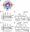
Instability of the H3T nucleosome. (A) H3T nucleosomes, reconstituted using 1.2 mg/mL total histones and 0.7 mg/mL DNA, were analyzed by nondenaturing 6% PAGE. Lane 1 indicates naked DNA. Lanes 2 and 3 indicate the H3T nucleosomes before and after a 55 °C incubation, respectively. DNA was visualized by ethidium bromide staining. Asterisks indicate bands corresponding to nonnucleosomal DNA–histone complexes. (B) The H3T and H3.1 nucleosomes were purified using a Prepcell apparatus, and were analyzed by nondenaturing 6% PAGE with ethidium bromide staining. (C) Histone compositions of the purified H3T and H3.1 nucleosomes were analyzed by 18% SDS-PAGE with Coomassie brilliant blue staining. (D) Salt titration. The nucleosomes were incubated in the presence of 0.4 M (lanes 1 and 5), 0.6 M (lanes 2 and 6), 0.7 M (lanes 3 and 7), and 0.8 M NaCl (lanes 4 and 8) at 42 °C for 2 h. The samples were analyzed by nondenaturing 6% PAGE with ethidium bromide staining. Lanes 1–4 and 5–8 indicate experiments with H3.1 and H3T nucleosomes, respectively. Bands corresponding to nucleosome monomers and nucleosome-nucleosome aggregates are indicated. Asterisks represent bands corresponding to nonnucleosomal DNA-histone complexes.
Fig. 2.
Fig. 2.
H2A/H2B associates weakly with H3T/H4. (A) The crystal structure of the H3.1 nucleosome determined in this study. Locations the PstI and EcoRI sites are indicated. The H2A/H2B and H3.1/H4 molecules are colored in purple and in dark blue, respectively. (B and C) H2A/H2B disassembly assay with hNap1. The nucleosomes were treated with PstI or EcoRI in the presence or absence of excess amount of hNap1 (6.5 μM). The resulting DNA fragments were extracted by Phenol/chloroform, and were analyzed by 10% PAGE with ethidium bromide staining. Arrows indicate the DNA fragment produced by complete PstI digestion. These results were confirmed to be reproduced in three independent experiments. (B) The H3T nucleosome. (C) The H3.1 nucleosome. (D and E) Interaction between H2A/H2B and H3T/H4 or H3.1/H4. H2A, H2B, H4, and H3T (D) or H3.1 (E) were incubated without DNA in the presence of 2 M NaCl. The samples were then subjected to HiLoad 26/60 Superdex 200 prep grade gel filtration column chromatography. Histone compositions of the peak fractions were analyzed by 18% SDS-PAGE with Coomassie brilliant blue staining. The peak fractions denoted as a, b, and c correspond to H2A/H2B/H3/H4 octamer, H3/H4 tetramer, and H2A/H2B dimer, respectively.
Fig. 3.
Fig. 3.
FRAP. HeLa cells expressing GFP-H3.1 or GFP-H3T were subjected to the FRAP analysis. (A) The mobility of GFP-H3.1 or GFP-H3T in living cells was analyzed by bleaching one-half of the nucleus. (B) The averages of the relative fluorescence intensity of bleached area were plotted with the standard deviations (n = 5). (C) GFP-H3T was incorporated into the HeLa cell chromatin. (Upper ) DNA fragments of mono-, di-, and trinucleosomes fractionated by sucrose gradient centrifugation were analyzed by agarose gel electrophoresis with ethidium bromide staining. (Right and Left) The nucleosome samples from the HeLa cells with and without GFP-H3T expression, respectively. The sucrose gradient fraction numbers are indicated at the top of each panel. Middle panel. Histone compositions of the purified nucleosomes were analyzed by 16% SDS-PAGE with Coomassie brilliant blue staining staining. (Lower) GFP-H3T was detected with anti-GFP monoclonal antibody.
Fig. 4.
Fig. 4.
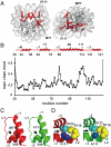
Crystal structure of the H3T nucleosome. (A) Two views of the H3T-nucleosome structure are represented. The H3T molecules are shown in red. Locations of the Met71 and Val111 residues are indicated. (B) Structural differences between H3T and H3.1 in the nucleosomes. The H3T and H3.1 structures are superimposed, and the rmsd values for each residue pair is calculated and plotted. The secondary structure of H3T in the nucleosome is shown in the top of the panel. Arrows indicate the locations of the H3T-specific amino acid residues, Met71, Ser98, and Val111. (C and D) Comparison of the H3T structure (red) with the H3.1 structure (green). The side chains of the H3T-M71, H3.1-V71, H3T-V89, H3.1-V89, H3T-V111, H3.1-A111, H3T-R116, H3.1-R116, H3T-D123, and H3.1-D123 residues are represented by space-filling models. The H3T and H3.1 regions containing the amino acid residues 71 (C) and 111 (D) are shown. Arrows in C and D indicate the locations of H3T and H3.1 that are structurally different from each other.
Fig. 5.
Fig. 5.
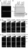
Mutational analysis of H3T and H3.1 nucleosomes. (A) Nucleosomes containing H3T and H3.1 mutants were purified using a Prepcell apparatus and were analyzed by nondenaturing 6% PAGE with ethidium bromide staining. (B) Histone compositions of the purified nucleosomes containing H3T and H3.1 mutants were analyzed by 18% SDS-PAGE with Coomassie brilliant blue staining. (C and D) Salt titration. Nucleosomes were incubated in the presence of 0.4 M (lanes 1, 5, 9, 13, and 17), 0.6 M (lanes 2, 6, 10, 14, and 18), 0.7 M (lanes 3, 7, 11, 15, and 19), and 0.8 M NaCl (lanes 4, 8, 12, 16, and 20) at 42 °C for 2 h. The samples were analyzed by nondenaturing 6% PAGE with ethidium bromide staining. Bands corresponding to nucleosome monomers and nucleosome–nucleosome aggregates are indicated. Asterisks represent bands corresponding to nonnucleosomal DNA-histone complexes. (C) The stability of H3T mutants. Lanes: 1–4, H3T; 5–8, H3T-M71V; 9–12, H3T-S98A; 13–16, H3T-V111A; and 17–20, H3T-M71V/V111A nucleosomes. (D) The stability of H3.1 mutants. Lanes: 1–4, H3.1; 5–8, H3.1-V71M; 9–12, H3.1-A98S; 13–16, H3.1-A111V; and 17–20, H3.1-V71M/A111V.
Fig. 6.
Fig. 6.
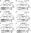
Mutational analyses of interactions of H2A/H2B with H3T/H4 or H3.1/H4. Gel filtration analyses were performed as described in Fig. 2 D and E, except that a HiLoad 16/60 Superdex 200 prep grade column was used. Fractions indicated with dots are analyzed by 18% SDS-PAGE with Coomassie brilliant blue staining. The peak fractions denoted as a, b, and c correspond to H2A/H2B/H3/H4 octamer, H3/H4 tetramer, and H2A/H2B dimer, respectively. (A) H3.1-V71M. (B) H3.1-A98S. (C) H3.1-A111V. (D) H3T-M71V. (E) H3T-S98A. (F) H3T-V111A.

References

    1. Govin J, Caron C, Lestrat C, Rousseaux S, Khochbin S. The role of histones in chromatin remodelling during mammalian spermiogenesis. Eur J Biochem. 2004;271:3459–3469. - PubMed
    1. Hammoud SS, et al. Distinctive chromatin in human sperm packages genes for embryo development. Nature. 2009;460:473–478. - PMC - PubMed
    1. Franklin SG, Zweidler A. Non-allelic variants of histones 2a, 2b and 3 in mammals. Nature. 1977;266:273–275. - PubMed
    1. Albig W, Ebentheuer J, Klobeck G, Kunz J, Doenecke D. A solitary human H3 histone gene on chromosome 1. Hum Genet. 1996;97:486–491. - PubMed
    1. Witt O, Albig W, Doenecke D. Testis-specific expression of a novel human H3 histone gene. Exp Cell Res. 1996;229:301–306. - PubMed

Publication types