. 2010 Aug;31(8):1456-64.
doi: 10.1093/carcin/bgq100. Epub 2010 May 24.

An unusual function of RON receptor tyrosine kinase as a transcriptional regulator in cooperation with EGFR in human cancer cells

Affiliations

An unusual function of RON receptor tyrosine kinase as a transcriptional regulator in cooperation with EGFR in human cancer cells

Hsiao-Sheng Liu et al. Carcinogenesis. 2010 Aug.

Abstract

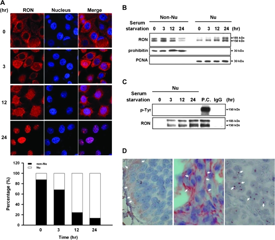
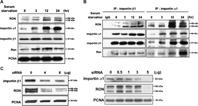
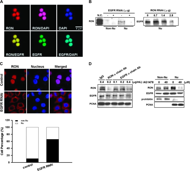
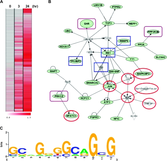
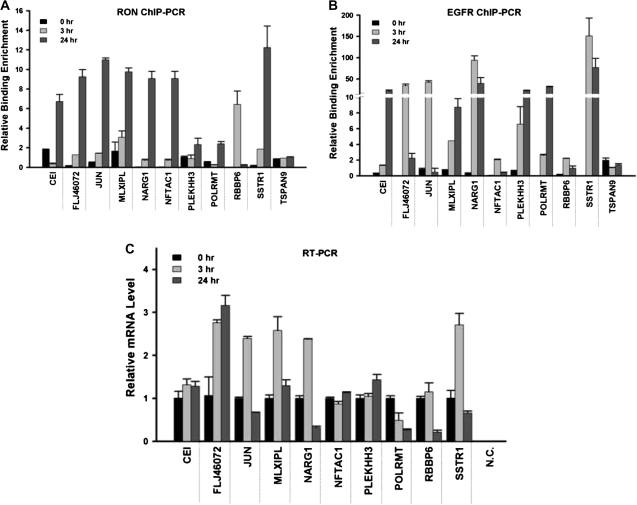
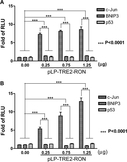
Homodimerization of RON (MST1R), a receptor tyrosine kinase, usually occurs in cells stimulated by a ligand and leads to the downstream activation of signaling pathways. Here we report that bladder cancer cells, in response to physiological stress, use an alternative mechanism for signaling activation. Time-course studies indicated that RON migrated directly from the membrane to the nucleus of bladder cancer cells in response to serum starvation. Biochemical and genetic studies implied that this nuclear internalization was complexed with epidermal growth factor receptor (EGFR) and required the docking of importins. In vivo analysis confirmed that nuclear RON was present in 38.4% (28/73) of primary bladder tumors. Chromatin immunoprecipitation (ChIP) on microarray analysis further revealed that this internalized complex bound to at least 134 target genes known to participate in three stress-responsive networks: p53, stress-activated protein kinase/c-jun N-terminal kinase and phosphatidylinositol 3-kinase/Akt. These findings suggest that RON, in a complex with EGFR, acts as a transcriptional regulator in response to acute disturbances (e.g. serum starvation) imposed on cancer cells. In an attempt to re-establish homeostasis, these cells bypass regular mechanisms required by ligand stimulation and trigger the RON-directed transcriptional response, which confers a survival advantage.


Figures

Fig. 1.
Fig. 1.
Kinetic analysis of nuclear localization of RON. TSGH8301 cells were serum starved for different time periods from 0, 3, 12 to 24 h. (A) Immunofluorescence analysis was conducted to monitor the time-dependent nuclear localization of RON. Treated cells were incubated with anti-RON (red) and then a Rhodamine-conjugated secondary antibody (upper panel). Nuclei were stained with 4′,6-diamidino-2-phenylindole (blue). The purple signals (merged) highlight the localization of RON in the nucleus. The results were quantified and expressed as a histogram (bottom panel). (B) Western blotting with anti-RON antibody on subcellular fractions of treated cells was exhibited to determine time-dependent nuclear import of RON. The prohibitin and proliferating cell nuclear antigen (PCNA) were chosen as markers for non-nucleus and nucleus fractions, respectively. (C) Nuclear fractions from treated cells were subjected to co-immunoprecipitation analysis with anti-RON antibody and then probed with anti-tyrosine (p-Tyr) antibody. Total cell lysate with MSP treatment was used as positive control (P.C.) for phospho-RON staining, and IgG was used as negative control of immunoprecipitation assay. (D) Immunohistochemical analysis was performed to detect RON expression on human bladder tissues from bladder cancer patients (n = 73). In this representative example, white arrows indicate positive staining areas of RON. Both ‘right’ and ‘middle panels’ are malignant bladder tissues; the ‘left panel’ is normal bladder tissue.
Fig. 2.
Fig. 2.
Importins/NLS-mediated nuclear import of RON. TSGH8301 cells were starved for different time periods as indicated and then nuclear extracts were collected for the following western blotting and immunoprecipitation assays with the indicated antibodies. (A) Upon serum starvation, increasing expression of RON, importin α1, importin β1 and Ran in the nucleus were detected by immunoblotting analysis. Proliferating cell nuclear antigen (PCNA) was used as a nuclear marker. (B) The association of RON with nuclear importin proteins, including importin α1, importin β1 and Ran, was determined by co-immunoprecipitation assays. Nuclear extracts were pulled down by either anti-importin β1 (left) or anti-importin α1 (right) antibodies and then probed with the indicated antibodies. (C) Inhibition of nuclear RON expression by importin α1 or β1 siRNA knockdown. TSGH8301 cells were transfected with different dosage of importin α1 or β1 siRNA for 48 h and then nuclear extracts were collected for sodium dodecyl sulfate–polyacrylamide gel electrophoresis resolution. PCNA was as a nuclear marker and loading control. A dose-dependent suppressor effect was more apparent in knock down of importin β1.
Fig. 3.
Fig. 3.
Interactions between EGFR and nuclear RON in bladder cancer cells. (A) Immunofluorescence analysis showed that, upon 24 h of serum starvation, RON (red) was colocalized with EGFR (green) in the nucleus of TSFG8301 cells. Nuclei were counterstained with 4′,6-diamidino-2-phenylindole (DAPI) (blue) and images acquired in an Olympus Confocal Microscope. (B) Nuclear RON expression was reduced by EGFR siRNA in TSGH8301 cells. After 48 h of either EGFR (left) or RON (right) siRNA transfection, nuclear extracts were collected for immunoblotting analysis. (C) RON (red) location was redistributed upon attenuating EGFR expression by EGFR siRNA. After 48 h transfection of EGFR siRNA, TSGH8301 cells were subjected to immunofluorescence analysis. Nuclei were counterstained with DAPI (blue). Staining intensities were quantified and expressed as histogram (bottom panel). (D) TSGH8301 cells were treated with indicated doses of EGFR neutralization antibody (left panel; lanes 3–5) and nuclear extracts were collected for western blot analysis. Treatment of RON α chain antibody (lane 3) was utilized as a positive control, whereas normal IgG antibody (lane 1) was used as a negative control to the neutralization antibodies. In ‘right panel’, cells were pretreated with optimized doses of AG1478, an EGFR-specific inhibitor, and then serum-starved for 24 h. Subcellular fractions were collected for immunoblotting with antibodies as indicated.
Fig. 4.
Fig. 4.
Identification of RON-targeting genes. (A) ChIP assay coupled with microarray (ChIP-chip) was conducted to identify the potential targets of RON, which is summarized into a heat map of 134 genes. Starved TSGH8301 cells at the indicated time periods (0, 3 and 24 h) were harvested for ChIP assay with anti-RON antibody. The dye-coupled-immunoprecipitated DNA was hybridized onto the 185K CGI microarrays. (B) Nuclear RON targets form a network of interactions by Ingenuity Pathway Analysis software. The input was all 134 significant genes from ChIP-chip assay. Genes depicted in green are the RON-targeting genes. Targets in red circles are associated with the stress-activated protein kinase/JNK signaling pathway, targets in blue rectangles are related to the p53 signaling pathways and targets in purple rectangles are associated with the phosphatidylinositol 3-kinase/Akt pathway. (C) The motif logo of consensus sequence to RON-targeting genes. The potential nuclear RON-targeting genes with ChIP–PCR validation were collected to establish a dataset for the motif analysis. The putative sequence was determined by MDscan (Motif Discovery Scan; http://ai.stanford.edu/∼xsliu/MDscan). The motif log was generated on http:weblogo.berkeley.edu.
Fig. 5.
Fig. 5.
Quantitative PCR analyses of candidate target genes. Quantitative ChIP–PCR analyses were conducted to validate the potential target genes of nuclear RON. After serum starvation for the indicated time periods, TSGH8301 cells were harvested and ChIP assays were performed with antibodies directed against RON (A) or EGFR (B). The immunoprecipitation DNA corresponding to the interested targets was measured by quantitative PCR. Quantitation of specific RON targeting was determined as a percent of input DNA and each error bar represents standard deviation calculated from triplicates. (C) Quantitative real-time–PCR analysis was performed to investigate the expression level of candidate target genes. Each error bar represents standard deviation calculated from triplicates. Amplified samples without reverse transcription were used as negative controls.
Fig. 6.
Fig. 6.
Transcription activation of putative target genes of RON. The promoter activity in HEK293 cells co-transfected with the indicated target gene reporter plasmids together with RON plasmid at various dosages was determined. The means ± SDs (error bars) were derived from three independent experiments. The promoter activity represented by luciferase activity was analyzed by the Dual-Glo™ Luciferase Assay System at 0, 3, 24 and 48 h after transfection, respectively. The results at 3 and 24 h were shown in 6A and 6B, respectively (P < 0.0001, by two-way analysis of variance test). The promoter activity was shown as fold of RLU (relative to the level of control vector reporter gene after normalization with co-transfected Renilla luciferase activity). This experiment was conducted in triplicate. The promoter activity after EGF treatment was used as a positive control. The empty vector pRLTK was used as an internal control.

Similar articles

Cited by

References

    1. Schlessinger J. Cell signaling by receptor tyrosine kinases. Cell. 2000;103:211–225. - PubMed
    1. Li E, et al. Role of receptor tyrosine kinase transmembrane domains in cell signaling and human pathologies. Biochemistry. 2006;23:6241–6251. - PMC - PubMed
    1. Sorkin A, et al. Signal transduction and endocytosis: close encounters of many kinds. Nat. Rev. 2002;3:600–614. - PubMed
    1. Gschwind A, et al. Cell communication networks: epidermal growth factor receptor transactivation as the paradigm for inter-receptor signal transmission. Oncogene. 2001;20:1594–1600. - PubMed
    1. Ronsin C, et al. A novel putative receptor protein tyrosine kinase of the met family. Oncogene. 1993;8:1195–1202. - PubMed

Publication types