Review
. 2010 Sep 15;588(Pt 18):3365-73.
doi: 10.1113/jphysiol.2010.190769. Epub 2010 May 24.

Kinase regulation of Na+-K+-2Cl- cotransport in primary afferent neurons

Affiliations
Review

Kinase regulation of Na+-K+-2Cl- cotransport in primary afferent neurons

Eric Delpire et al. J Physiol. .

Abstract

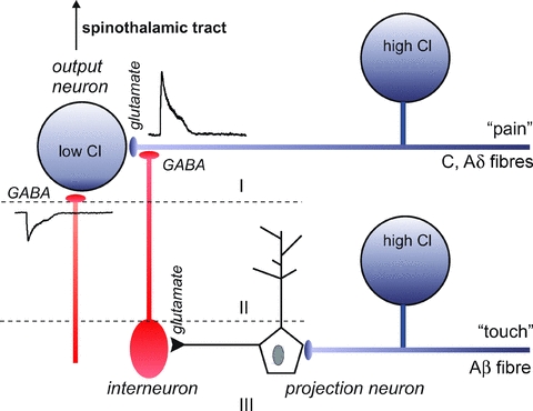
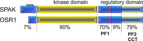
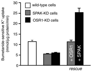
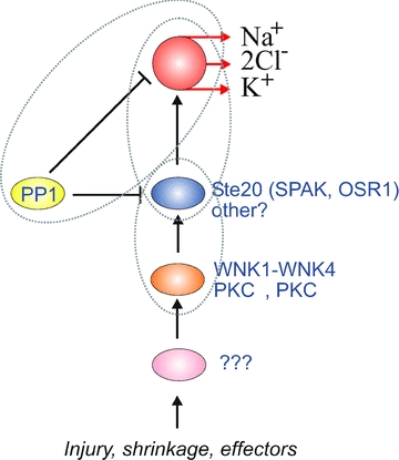
The Na(+)-K(+)-2Cl(-) cotransporter NKCC1 is expressed in sensory neurons where it accumulates intracellular Cl(-) and facilitates primary afferent depolarization. Depolarization of primary afferent fibre terminals interferes with the gating of incoming sensory signals to the spinal cord. The cotransporter belongs to a family of ion transporters which are sensitive to changes in cell volume. Cell shrinkage, through mechanisms that are still unknown, leads to the phosphorylation and activation of NKCC1. Similarly, axotomy results in increased NKCC1 phosphorylation in dorsal root ganglion (DRG) neurons. This review summarizes the work on the kinases that directly mediate NKCC1 activation. These are the sterile-20-like kinases SPAK and OSR1. Upon their activation through phosphorylation by upstream kinases, SPAK and OSR1 bind to specific peptides located in the cytosolic N-terminal tail of NKCC1, phosphorylate, and stimulate cotransport activity. Expression of SPAK and OSR1 varies from tissue to tissue, but in DRG neurons and in spinal cord, SPAK and OSR1 expression levels are similar. In DRG neurons, both kinases participate in the modulation of NKCC1, as the knockdown of one kinase only results in a partial decrease of NKCC1 function, while the knockdown of both kinases is additive. The identity of the kinases (e.g. WNK kinases) that possibly act upstream of SPAK and OSR1 is also discussed.


Figures

Figure 1
Figure 1. Schematic diagram of adult CNS neuron with low intracellular Cl and immature CNS neuron or adult sensory neuron with high intracellular Cl
Opening of GABAA receptor in the membrane leads to Cl entry and hyperpolarization in the first neuron, and Cl exit and depolarization in the second neuron.
Figure 2
Figure 2. Schematic representation of the pain circuitry in the spinal cord
Unmyelinated (C) and thinly myelinated (Aδ) afferent fibres bring pain signals to output neurons in lamina I and II in the spinal cord. Interneurons activated by projection neurons in deeper layers release GABA at the terminals of the C and Aδ fibres. As the Cl concentration is high in these afferent neurons, GABA produces depolarization of the terminal. This depolarization inhibits incoming pain signals coming from the periphery.
Figure 3
Figure 3. Schematic representation of SPAK and OSR1 kinases with short N-terminal domain, followed by catalytic domain, and large C-terminal regulatory domain
Numbers represent the percentage amino acid identity between mouse SPAK and OSR1 sequences within the delimited region. Red lines indicate the position of a putative caspase cleavage site. PAPA = Proline Alanine rich region.
Figure 4
Figure 4. NKCC1 activity, as measured through bumetanide-sensitive K+ uptake, in wild-type and SPAK- or OSR1-knockdown 50B11 cells
Note the reduction in activity in both knockdown cell lines. OSR1 overexpression was able to restore NKCC1 activity in SPAK-knockdown cells. Similarly, SPAK over-expression was able to activate NKCC1 in OSR1-knockdown cells. Redrawn from Geng et al. (2009).
Figure 5
Figure 5. Illustration of different regulatory elements of NKCC1
SPAK and OSR1 represent kinases that are directly responsible for phosphorylation and activation of NKCC1. Both NKCC1 and these direct kinases are inactivated through dephosphorylation by PP1. WNK kinases and several PKC isotypes are located upstream of SPAK and OSR1. These kinases are also probably the target of other unidentified effectors. Venn diagrams represent necessary protein–protein interaction between regulatory elements.
None

References

    1. Alvarez-Leefmans FJ. Chloride transporters in presynaptic inhibition, pain and neurogenic inflammation. In: Alvarez-Leefmans FJ, Delpire E, editors. Physiology and Pathology of Chloride Transporter and Channels in the Nervous System: From Molecules to Diseases. London: Academic Press; 2009. pp. 439–470.
    1. Alvarez-Leefmans FJ, Gamiño SM, Giraldez F, Nogueron I. Intracellular chloride regulation in amphibian dorsal root ganglion neurones studied with ion-selective microelectrodes. J Physiol. 1988;406:225–246. - PMC - PubMed
    1. Anselmo AN, Earnest S, Chen W, Juang YC, Kim SC, Zhao Y, Cobb MH. WNK1 and OSR1 regulate the Na+, K+, 2Cl− cotransporter in HeLa cells. Proc Natl Acad Sci U S A. 2006;103:10883–10888. - PMC - PubMed
    1. Blaesse P, Airaksinen MS, Rivera C, Kaila K. Cation-chloride cotransporters and neuronal function. Neuron. 2009;61:820–838. - PubMed
    1. Cervero F, Laird JM. Mechanisms of allodynia: interactions between sensitive mechanoreceptors and nociceptors. Neuroreport. 1996;7:526–528. - PubMed

Publication types

MeSH terms

Substances

LinkOut - more resources