

Peyer's patch-deficient mice demonstrate that Mycobacterium avium subsp. paratuberculosis translocates across the mucosal barrier via both M cells and enterocytes but has inefficient dissemination
- PMID: 20498259
- PMCID: PMC2916292
- DOI: 10.1128/IAI.01411-09
Peyer's patch-deficient mice demonstrate that Mycobacterium avium subsp. paratuberculosis translocates across the mucosal barrier via both M cells and enterocytes but has inefficient dissemination
Abstract
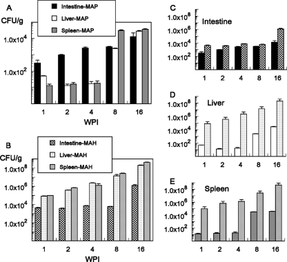
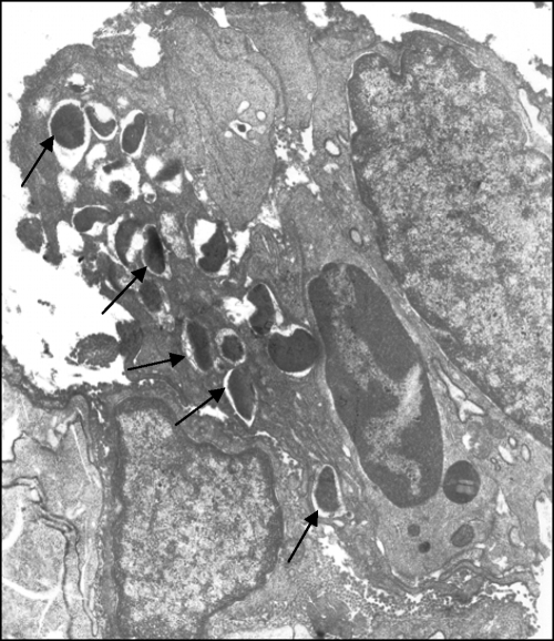
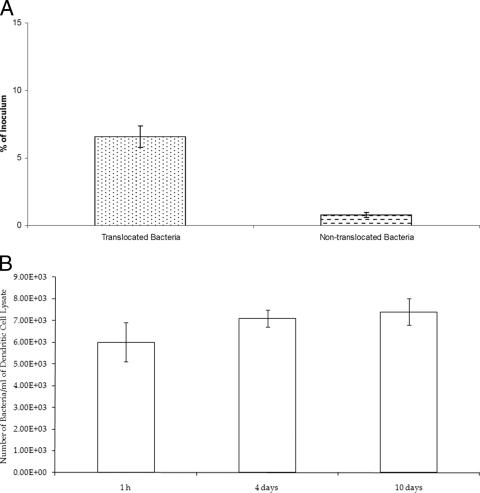
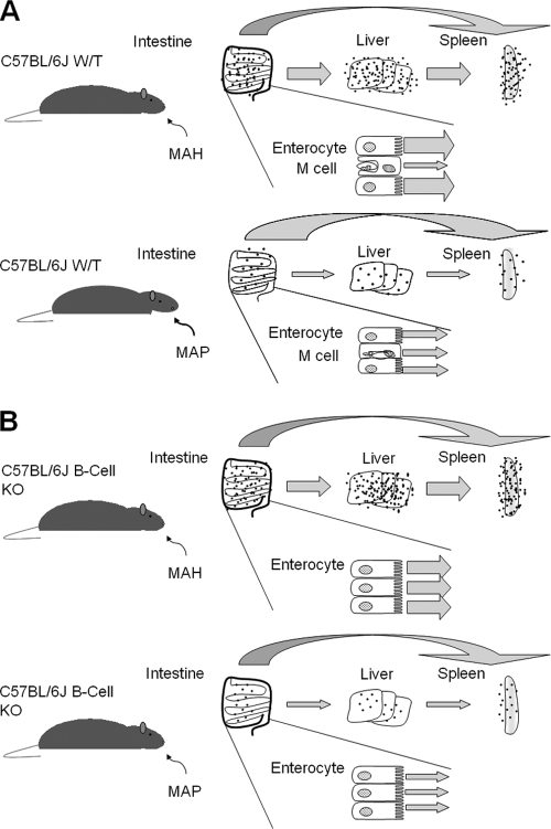
Mycobacterium avium subsp. paratuberculosis, the agent of Johne's disease, infects ruminant hosts by translocation through the intestinal mucosa. A number of studies have suggested that M. avium subsp. paratuberculosis interacts with M cells in the Peyer's patches of the small intestine. The invasion of the intestinal mucosa by M. avium subsp. paratuberculosis and Mycobacterium avium subsp. hominissuis, a pathogen known to interact with intestinal cells, was compared. M. avium subsp. paratuberculosis was capable of invading the mucosa, but it was significantly less efficient at dissemination than M. avium subsp. hominissuis. B-cell knockout (KO) mice, which lack Peyer's patches, were used to demonstrate that M. avium subsp. paratuberculosis enters the intestinal mucosa through enterocytes in the absence of M cells. In addition, the results indicated that M. avium subsp. paratuberculosis had equal abilities to cross the mucosa in both Peyer's patch and non-Peyer's patch segments of normal mice. M. avium subsp. paratuberculosis was also shown to interact with epithelial cells by an alpha(5)beta(1) integrin-independent pathway. Upon translocation, dendritic cells ingest M. avium subsp. paratuberculosis, but this process does not lead to efficient dissemination of the infection. In summary, M. avium subsp. paratuberculosis interacts with the intestinal mucosa by crossing both Peyer's patches and non-Peyer's patch areas but does not translocate or disseminate efficiently.
Figures
References
-
- Alonso-Hearn, M., D. Patel, L. Danelishvili, L. Meunier-Goddik, and L. E. Bermudez. 2008. The Mycobacterium avium subsp. paratuberculosis MAP3464 gene encodes an oxidoreductase involved in invasion of bovine epithelial cells through the activation of host cell Cdc42. Infect. Immun. 76:170-178. - PMC - PubMed
-
- Bermudez, L. E., M. Petrofsky, P. Kolonoski, and L. S. Young. 1992. An animal model of Mycobacterium avium complex disseminated infection after colonization of the intestinal tract. J. Infect. Dis. 165:75-79. - PubMed
-
- Bermudez, L. E., F. J. Sangari, P. Kolonoski, M. Petrofsky, and J. Goodman. 2002. The efficiency of the translocation of Mycobacterium tuberculosis across a bilayer of epithelial and endothelial cells as a model of the alveolar wall is a consequence of transport within mononuclear phagocytes and invasion of alveolar epithelial cells. Infect. Immun. 70:140-146. - PMC - PubMed
-
- Chacon, O., L. E. Bermudez, and R. G. Barletta. 2004. Johne's disease, inflammatory bowel disease, and Mycobacterium paratuberculosis. Annu. Rev. Microbiol. 58:329-363. - PubMed
Publication types
MeSH terms
LinkOut - more resources
Full Text Sources
Molecular Biology Databases
Research Materials
