. 2010 Aug;78(8):3484-92.
doi: 10.1128/IAI.00117-10. Epub 2010 May 24.

CD4 T-cell suppression by cells from Toxoplasma gondii-infected retinas is mediated by surface protein PD-L1

Affiliations

CD4 T-cell suppression by cells from Toxoplasma gondii-infected retinas is mediated by surface protein PD-L1

Elizabeth Charles et al. Infect Immun. 2010 Aug.

Abstract

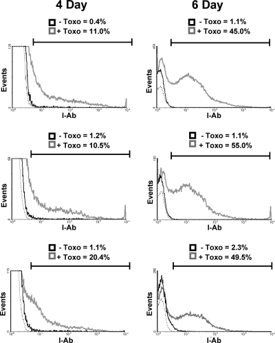
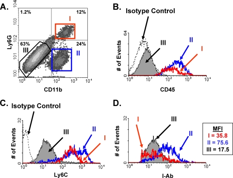
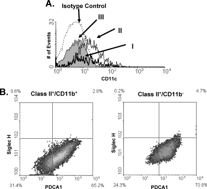
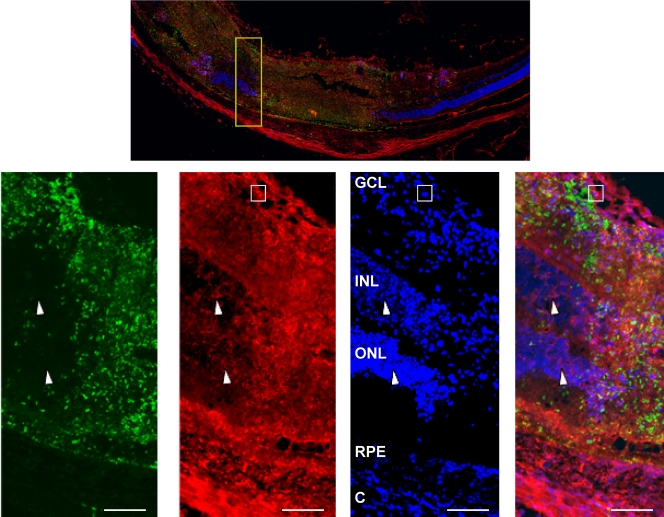
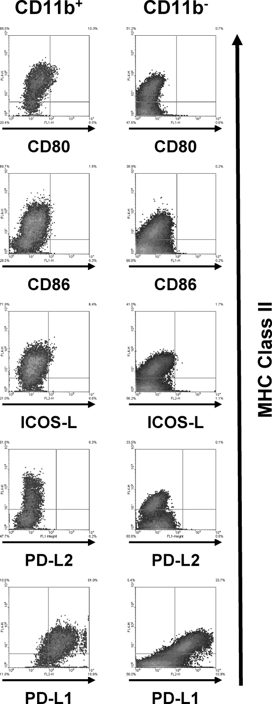
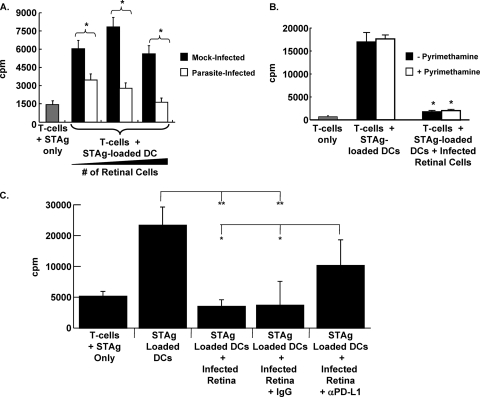
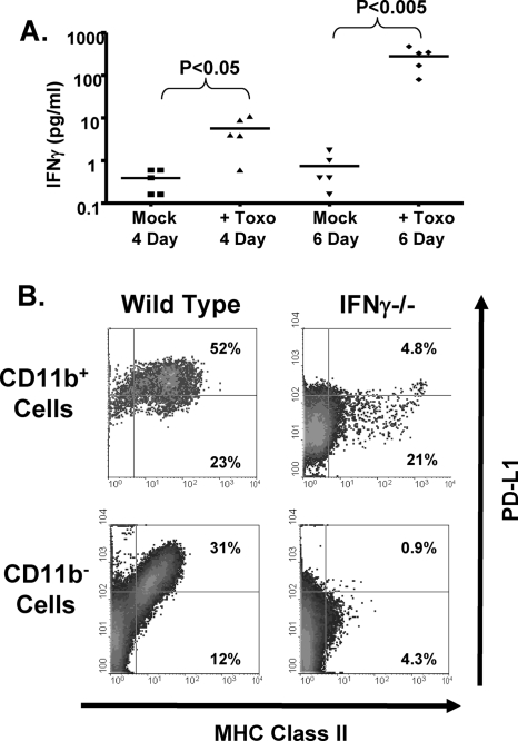
In the inflamed retina, CD4(+) T cells can cause retinal damage when they are not properly regulated. Since tissue expression of major histocompatibility complex (MHC) class II and costimulatory molecules is a key mechanism for regulating effector T cells, we tested the hypothesis that upregulation of these proteins in the retina contributes to the regulation of CD4 T cells. Here we report that in retinas infected with the protozoan parasite Toxoplasma gondii, MHC class II is upregulated on infiltrating leukocytes as well as on resident retinal cells, including photoreceptors. Flow cytometric analysis indicated that B7 costimulatory family members (CD80, CD86, ICOS-L, and programmed death ligand 2 [PD-L2]) were not expressed on class II(+) cells. In contrast, PD-L1 (also named B7-H1 or CD274) was expressed on the majority of both hematopoietic and resident retinal MHC class II-expressing cells. Retinal cells from Toxoplasma-infected animals were able to suppress T-cell activation in a PD-L1-dependent manner. Finally, we demonstrate that the expression of MHC class II and PD-L1 was critically dependent on gamma interferon (IFN-gamma) expression. These data suggest that retinal MHC class II and PD-L1 expression is a novel mechanism by which the retina protects itself from CD4 T-cell-mediated immune damage in ocular toxoplasmosis and other types of retinal immune responses.


Figures

FIG. 1.
FIG. 1.
MHC class II expression in Toxoplasma-infected eyes. Mice were either mock infected (solid black lines) or intravitreally injected (gray lines) with 104 parasites. Four and 6 days later, eyes were harvested and analyzed by flow cytometry for MHC class II expression. The gate includes all MHC class II+ events as determined by isotype staining controls (dashed line). The y axes of the 4-day histograms were decreased to highlight class II+ cells. Shown are three representative histograms from independent experiments.
FIG. 2.
FIG. 2.
Three distinct populations of MHC class II-expressing cells in Toxoplasma-infected eyes. Mice were intravitreally injected with 104 parasites, and 6 days later, eyes were harvested and analyzed by using flow cytometry. (A) FACS plot of CD11b and Ly6G expression on MHC class II+-gated cells. The distinct populations are highlighted as I (CD11b+ Ly6G+), II (CD11b+ Ly6G), and III (CD11b Ly6G). (B through D) Histograms of CD45 (B), Ly6C (C), and MHC class II (D) expression levels in each of the 3 populations. The dashed lines in panels B and C represent isotype control staining levels. MFI, mean fluorescence intensity of each population. Representative results from three independent experiments are shown.
FIG. 3.
FIG. 3.
Dendritic cell staining in Toxoplasma-infected eyes. Mice were intravitreally injected with 104 parasites. Six days later, eyes were harvested and analyzed using flow cytometry. (A) Histogram of CD11c expression on the three populations of MHC class II-expressing cells from Fig. 2A. (B) FACS plots of PDCA1 and Siglec-H staining of class II+ CD11b+ and class II+ CD11b cells.
FIG. 4.
FIG. 4.
MHC class II is expressed on resident retinal cells in Toxoplasma-infected retinas. Mice were intravitreally injected with 104 parasites. Six days later, eyes were harvested, fixed, and processed for immunocytochemistry. Sections were stained to detect either DAPI (blue), MHC class II (red), or SAG1 (green). (Top) Low-magnification (×40) image of the posterior retina from an infected eye. (Bottom) High-magnification (×400) images of the area highlighted in the yellow box in the top image. Note the SAG1 staining and the intense class II staining in necrotic regions of the retina. Arrowheads point to class II+ nuclear cells in inner and outer nuclear layers. The boxes highlight a crescent-shaped, class IIhi monocyte. Bars, 100 μm. Abbreviations: C, choroid; RPE, retinal pigmented epithelium; ONL, outer nuclear layer; INL, inner nuclear layer; GCL, ganglion cell layer.
FIG. 5.
FIG. 5.
B7 family member expression on MHC class II+ cells. Mice were intravitreally injected with 104 parasites. Six days later, eyes were harvested and stained with anti-MHC class II and the indicated antibodies. Representative FACS plots from three independent experiments are shown.
FIG. 6.
FIG. 6.
T-cell suppression by retinal cells from Toxoplasma-infected retinas is PD-L1 dependent. (A) Splenic T cells isolated from cpsII-vaccinated mice were incubated with STAg alone (shaded bar) or STAg-loaded splenic APCs in the presence of increasing numbers (1 × 10⁵, 5 × 10⁴, or 1 × 10⁴) of retinal cells from either mock-infected (filled bars) or parasite-infected (open bars) mice. Shown are the averages for quadruplicate samples repeated two independent times. *, P < 0.005 by a two-tailed, unpaired Student t test. (B) Splenic T cells from vaccinated mice were incubated with STAg alone (shaded bar) or with STAg-loaded DCs in the absence or presence of parasite-infected retinal cells. Pyrimethamine (1 μM) was included as indicated. Shown are results of a representative experiment repeated three times in quadruplicate. *, P < 0.005 by Student's t test. (C) Splenic T cells from vaccinated mice were incubated with the indicated cells (5 × 10⁴ parasite-infected retinal cells were used where indicated) and antibodies. Shown are results of a representative experiment carried out in quadruplicate. Asterisks indicate significance by Student's t test (**, P < 0.01; *, P < 0.05).
FIG. 7.
FIG. 7.
IFN-γ is important for MHC class II and PD-L1 expression in Toxoplasma-infected eyes. (A) Mice were intravitreally injected with 104 parasites (Toxo). After 4 and 6 days, eyes were harvested and enucleated, and IFN-γ levels were measured in lysates prepared from posterior segments. P values were determined using an unpaired, nonparametric Student t test. (B) Wild-type and IFN-γKO mice were intravitreally injected with 104 parasites. Six days later, eyes were harvested and processed for flow cytometry. Shown are representative FACS plots of MHC class II and PD-L1 expression from three independent experiments.

Similar articles

Cited by

References

    1. Blasius, A. L., M. Cella, J. Maldonado, T. Takai, and M. Colonna. 2006. Siglec-H is an IPC-specific receptor that modulates type I IFN secretion through DAP12. Blood 107:2474-2476. - PMC - PubMed
    1. Blasius, A. L., E. Giurisato, M. Cella, R. D. Schreiber, A. S. Shaw, and M. Colonna. 2006. Bone marrow stromal cell antigen 2 is a specific marker of type I IFN-producing cells in the naive mouse, but a promiscuous cell surface antigen following IFN stimulation. J. Immunol. 177:3260-3265. - PubMed
    1. Bliss, S. K., B. A. Butcher, and E. Y. Denkers. 2000. Rapid recruitment of neutrophils containing prestored IL-12 during microbial infection. J. Immunol. 165:4515-4521. - PubMed
    1. Brandt, C., P. Knupfer, G. Boush, R. Gausas, and J. Chandler. 1990. In vivo induction of Ia expression in murine cornea after intravitreal injection of interferon-gamma. Invest. Ophthalmol. Vis. Sci. 31:2248-2253. - PubMed
    1. Brooks, D. G., S. J. Ha, H. Elsaesser, A. H. Sharpe, G. J. Freeman, and M. B. Oldstone. 2008. IL-10 and PD-L1 operate through distinct pathways to suppress T-cell activity during persistent viral infection. Proc. Natl. Acad. Sci. U. S. A. 105:20428-20433. - PMC - PubMed

Publication types

LinkOut - more resources