. 2010 Aug;30(15):3875-86.
doi: 10.1128/MCB.00169-10. Epub 2010 May 24.

HuR uses AUF1 as a cofactor to promote p16INK4 mRNA decay

Affiliations

HuR uses AUF1 as a cofactor to promote p16INK4 mRNA decay

Na Chang et al. Mol Cell Biol. 2010 Aug.

Abstract

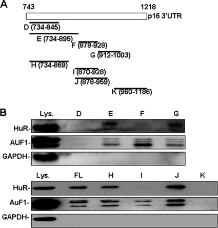
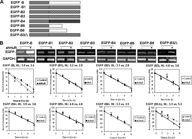
In this study, we show that HuR destabilizes p16(INK4) mRNA. Although the knockdown of HuR or AUF1 increased p16 expression, concomitant AUF1 and HuR knockdown had a much weaker effect. The knockdown of Ago2, a component of the RNA-induced silencing complex (RISC), stabilized p16 mRNA. The knockdown of HuR diminished the association of the p16 3' untranslated region (3'UTR) with AUF1 and vice versa. While the knockdown of HuR or AUF1 reduced the association of Ago2 with the p16 3'UTR, Ago2 knockdown had no influence on HuR or AUF1 binding to the p16 3'UTR. The use of EGFP-p16 chimeric reporter transcripts revealed that p16 mRNA decay depended on a stem-loop structure present in the p16 3'UTR, as HuR and AUF1 destabilized EGFP-derived chimeric transcripts bearing wild-type sequences but not transcripts with mutations in the stem-loop structure. In senescent and HuR-silenced IDH4 human diploid fibroblasts, the EGFP-p16 3'UTR transcript was more stable. Our results suggest that HuR destabilizes p16 mRNA by recruiting the RISC, an effect that depends on the secondary structure of the p16 3'UTR and requires AUF1 as a cofactor.


Figures

FIG. 1.
FIG. 1.
HuR represses p16 by destabilizing the p16 mRNA. (A) Forty-eight hours after the transfection of HeLa cells with a vector expressing Flag-HuR (left) or the HuR shRNA (right), lysates were prepared to assess the levels of HuR, p16, and loading control GAPDH by Western blot analysis. (B) RNA isolated from cells described for panel A was subjected to RT-qPCR to assess the mRNA levels of p16 and loading control GAPDH. The Western blotting and RT-qPCR data are representatives of three or more experiments. Where indicated (bottom panels), the signals were quantified by densitometry and represented as means ± SD from three independent experiments. (C) Cells described for panel A were exposed to actinomycin D (2 μg/ml), whereupon the cellular RNA was isolated at the times indicated and subjected to real-time PCR to assess the half-life of p16 mRNA.
FIG. 2.
FIG. 2.
p16 3′UTR interacts with HuR and confers the responsiveness of EGFP-p16 3′UTR reporters to the ectopic modulation of HuR expression. (A) Upper panels, schematic representation of the p16 mRNA fragments prepared for biotin pulldown assays. Bottom panels, biotin pulldown assays using biotinylated fragments to detect bound HuR by Western blotting. A 5-μg aliquot of whole-cell lysate (Lys.) and bound GAPDH were included. (B) Upper panels, schematic representation of the EGFP-p16 fragment reporters. HeLa cells stably transfected with the plasmid pTet-Off were cotransfected with each of the EGFP reporters (as depicted in the upper panels) plus a vector expressing either control or HuR shRNA. Forty-eight hours later, RNA was prepared and subjected to RT-qPCR to monitor the levels of the EGFP-p16 chimeric transcripts, using GAPDH mRNA as a loading control (bottom panels). (C) The transfections in Fig. 2B (bottom panels) were exposed to doxycyclin (Dox, 2 μg/ml) to shut off the expression of the EGFP-p16 chimeric transcripts, whereupon the RNA was prepared at the times indicated and subjected to real-time qPCR to assess the half-lives of the EGFP-p16 chimeric transcripts.
FIG. 3.
FIG. 3.
Identification of the HuR interaction motif in the p16 3′UTR. (A) Schematic representation of the p16 mRNA, depicting fragments used for biotin pulldown assays. (B) Biotin pulldown assays using biotinylated fragments to detect bound cytoplasmic HuR and AUF1 by Western blotting. A 5-μg aliquot of whole-cell lysate (Lys.) and bound GAPDH were included as controls.
FIG. 4.
FIG. 4.
Analysis of the secondary structure of the HuR interaction motif in the p16 3′UTR. (A) Schematic representation of the secondary structure (SL1 and SL2), analyzed as described in Materials and Methods. (B) Schematic representation depicting the wild-type fragment B and its mutants (B1 to B6 and B3Δ). The mutated sites are marked by capital letters. (C) Biotin pulldown assays were performed using biotinylated fragments (B to B6 and B3Δ) to evaluate the influence of the AUUUA pentamer and the SL1 or SL2 structure in the association of HuR, AUF1, and Ago2 with the p16 3′UTR. A 5-μg aliquot of whole-cell lysate (Lys.) and bound GAPDH were included as controls.
FIG. 5.
FIG. 5.
Influence of the SL1 or SL2 structure in HuR-mediated destabilization of EGFP-B chimeric transcript. HeLa cells stably transfected with pTet-Off plasmid were cotransfected with each of the EGFP-derived reporters bearing fragments (B to B6 and B3Δ) and a vector expressing HuR or the control shRNA. (B) Forty-eight hours later, the levels and the half-lives of the chimeric transcripts were assessed by RT-qPCR (upper panels) and real-time qPCR (bottom panels), respectively, as described in Materials and Methods.
FIG. 6.
FIG. 6.
SL1 or SL2 structure within the B fragment is required for AUF1 to destabilize the EGFP-B chemeric transcript. HeLa cells stably transfected with the pTet-Off plasmid were further transfected with a vector expressing AUF1 or control shRNA along with each of the EGFP-derived reporter vectors (Fig. 2B and 5A). Twenty-four hours later, the levels of AUF1 were monitored by Western blot analysis (A) and the half-lives of the chimeiric transcripts were assessed as described in Materials and Methods (B).
FIG. 7.
FIG. 7.
HuR and AUF1 interact with the p16 3′UTR, recruit the RISC, and destabilize p16 mRNA in a cooperative manner. HeLa cells were individually transfected with a vector expressing HuR (lane 2) or AUF1 (lane 3) shRNA or cotransfected with both vectors (lane 4). (A) Forty-eight h after transfection, whole-cell lysate and RNA were prepared and subjected to Western blotting and RT-qPCR analysis to assess the protein and mRNA levels of p16. GAPDH served as a loading control. (B) HeLa cells were transfected with siRNA targeting Ago2 (siAgo2, +) or the control siRNA (siAgo2, −). Twenty-four hours later, whole-cell lysate and RNA were prepared and subjected to Western blotting and RT-qPCR to assess the protein and mRNA levels of p16. GAPDH served as a loading control. Where indicated, the Western blot and RT-qPCR signals were quantified by densitometry and are represented as means ± SD from three independent experiments. (C) The half-life of p16 mRNA was assessed from the transfections in Fig. 7B, as described in Materials and Methods. (D) HeLa cells were transfected with a vector expressing HuR (left panels) or AUF1 (right panels) shRNA. Forty-eight hours later, cytoplasmic extracts were prepared for biotin pulldown assays using biotinylated p16 fragments B and B4. The bound HuR, AUF1, and Ago2 in the pull-down materials were detected by Western blotting. A 5-μg aliquot of whole-cell lysate (Lys.), pulldown using CR fragment (Neg.), and bound GAPDH were included as controls. (E) Cytoplasmic extracts from cells described for panel A were subjected to biotin pulldown analysis using biotinylated p16 fragment B. The bound HuR, AUF1, and Ago2 in the pulldown materials were detected by Western blotting, as described for panel D. (F) HeLa cells were transfected with a vector expressing HuR (left panels) or AUF1 shRNA (right panels) along with pGL3-B reporter vector. Forty-eight hours later, UV cross-link RNP IP assays were performed using cytoplasmic extracts, as described in Materials and Methods. The AUF1- and Ago2-associated pGL3-B chimeric transcripts in HuR-silenced cells (left two panels) as well as the HuR- and Ago2-bound pGL3-B chimeric transcripts in AUF1-silenced cells (right two panels) were quantified by real-time PCR. Values represent means ± SD from three independent experiments.
FIG. 8.
FIG. 8.
Knockdown of Ago2 has no influence on the association of the p16 3′UTR with HuR or AUF1. (A) HeLa cells were transfected with the siRNA targeting Ago2 or the control siRNA. Twenty-four hours later, cytoplasmic extracts were prepared and subjected to biotin pulldown assays using biotinylated fragments B and B4. The bound Ago2, HuR, and AUF1 were detected by Western blotting as described in the legend of Fig. 7D. (B) HeLa cells were transfected with pGL3-B vector; 24 h later, cells were further transfected with the siRNA targeting Ago2 or the control siRNA and cultured for an additional 24 h. UV cross-link RNP IP assays were performed using cytoplasmic extracts, as described in Materials and Methods. The HuR- or AUF1-bound pGL3-B chimeric transcripts in cells with silenced Ago2 were tested by real-time qPCR. Values represent means ± SD from three independent experiments.
FIG. 9.
FIG. 9.
HuR and AUF1 competitively associated with the p21 3′UTR. The cytoplasmic extracts described in the legend to Fig. 7D were used for biotin pulldown assays using biotinylated p21 3′UTR. Bound HuR and AUF1 in HuR (A)- or AUF1 (B)-silenced cells were assessed by Western blotting. A 5-μg aliquot of whole-cell lysate (Lys.) and bound GAPDH were included as controls.
FIG. 10.
FIG. 10.
HuR, AUF1, and Ago2 interact in the cytoplasm in an RNA-dependent manner. The cytoplasmic extracts prepared from HeLa cells were incubated with RNase A or left untreated. IP assays were performed using HuR or AUF1 (A) or Ago2 (B) antibody. The IP materials then were subjected to Western blotting to detect the presence of HuR, AUF1, and Ago2. IP with IgG served as a negative control.
FIG. 11.
FIG. 11.
Knockdown of Dicer or Drosha has no influence on the association of the p16 3′UTR with HuR, AUF1, or Ago2. Twenty-four hours after the transfection of HeLa cells with pGL3-B, cells were further transfected with siRNAs targeting Dicer (A), Drosha (B), or control siRNA and cultured for an additional 24 h. (A) On the left, the levels of Dicer, Ago2, HuR, AUF1, and p16 were assessed by Western blot analysis of whole-cell lysates. GAPDH was used as a loading control. On the right, cytoplasmic extracts were prepared and subjected to biotin pulldown assays using biotinylated fragment B; bound Ago2, HuR, and AUF1 were detected by Western blotting, as described in the legend to Fig. 7D. (B) On the left, the levels of Drosha, Ago2, HuR, AUF1, and p16 were assessed by Western blot analysis using whole-cell lysates. GAPDH was used as a loading control. On the right, cytoplasmic extracts were prepared and subjected to biotin pulldown assays using biotinylated fragment B. The bound Ago2, HuR, and AUF1 were detected by Western blotting, as described in the legend to Fig. 7D. (C and D) HeLa cells were transfected with pGL3-B vector. Twenty-four hours later, cells were further transfected with siRNAs targeting Dicer (C) or Drosha (D) and cultured for an additional 24 h. UV cross-link RNP IP assays were performed using cytoplasmic extracts, as described in Materials and Methods. The HuR-, AUF1-, and Ago2-bound pGL3-B chimeric transcripts in cells silenced for Dicer (C) or Drosha (D) were tested by real-time qPCR. Values represent means ± SD from three independent experiments.
FIG. 12.
FIG. 12.
Fragment B of the p16 3′UTR is a HuR response element in replicative senescence. (A and B) IDH4 cells were transfected with EGFP-CR or EGFP-B reporter vector, whereupon the cells were cultured either in regular medium plus dexamethasone (1 μg/ml; Y) or in medium containing charcoal-stripped serum in the absence of dexamethasone (to induce cell senescence; S) for 3 days. (A) Whole-cell lysates then were prepared to assess the level of HuR and Ago2 as well as the loading control GAPDH by Western blotting. (B) The half-lives of the EGFP-CR or EGFP-B transcripts were determined as described in Materials and Methods. Values represent means ± SD from five independent experiments. (C and D) IDH4 cells were transfected with pTet-Off plasmid. Twenty-four hours later, cells were further cotransfected with a vector expressing HuR or the control shRNA as well as EGFP-CR or EGFP-B reporter vector and cultured for an additional 24 h. Whole-cell lysate was prepared and subjected to Western blotting to monitor the level of HuR and loading control GAPDH (C). The half-lives of EGFP-CR and EGFP-B transcripts were tested as described in Materials and Methods (D). (E and F) IDH4 cells were transfected with pTet-Off plasmid along with EGFP-CR or EGFP-B reporter vector. Twenty-four hours later, cells were further transfected with siRNA targeting Ago2 or the control siRNA and cultured for an additional 24 h. Ago2 and GAPDH protein levels (E) as well as the half-lives of EGFP-CR and EGFP-B transcripts (F) were tested as described for panels C and D. Values represent means ± SD from five independent experiments. Where indicated, the Western blotting signals were quantified by densitometry.

Similar articles

Cited by

References

    1. Abdelmohsen, K., R. Pullmann, Jr., A. Lal, H. H. Kim, S. Galban, X. Yang, J. D. Blethrow, M. Walker, J. Shubert, D. A. Gillespie, H. Furneaux, and M. Gorospe. 2007. Phosphorylation of HuR by Chk2 regulates SIRT1 expression. Mol. Cell 25:543-557. - PMC - PubMed
    1. Atasoy, U., J. Watson, D. Patel, and J. D. Keene. 1998. ELAV protein HuA (HuR) can redistribute between nucleus and cytoplasm and is upregulated during serum stimulation and T cell activation. J. Cell Sci. 111(Pt 21):3145-3156. - PubMed
    1. Brennan, C. M., I. E. Gallouzi, and J. A. Steitz. 2000. Protein ligands to HuR modulate its interaction with target mRNAs in vivo. J. Cell Biol. 151:1-14. - PMC - PubMed
    1. Chemnitz, J., D. Pieper, C. Grüttner, and J. Hauber. 2009. Phosphorylation of the HuR ligand APRIL by casein kinase 2 regulates CD83 expression. Eur. J. Immunol. 39:267-279. - PubMed
    1. Chi, S. W., J. B. Zang, A. Mele, and R. B. Darnell. 2009. Argonaute HITS-CLIP decodes microRNA-mRNA interaction maps. Nature 460:479-486. - PMC - PubMed

Publication types

MeSH terms

Substances

LinkOut - more resources