. 2010 Aug;30(15):3767-78.
doi: 10.1128/MCB.00899-09. Epub 2010 May 24.

c-Fos proteasomal degradation is activated by a default mechanism, and its regulation by NAD(P)H:quinone oxidoreductase 1 determines c-Fos serum response kinetics

Affiliations

c-Fos proteasomal degradation is activated by a default mechanism, and its regulation by NAD(P)H:quinone oxidoreductase 1 determines c-Fos serum response kinetics

Julia Adler et al. Mol Cell Biol. 2010 Aug.

Abstract

The short-lived proto-oncoprotein c-Fos is a component of the activator protein 1 (AP-1) transcription factor. A large region of c-Fos is intrinsically unstructured and susceptible to a recently characterized proteasomal ubiquitin-independent degradation (UID) pathway. UID is active by a default mechanism that is inhibited by NAD(P)H:quinone oxidoreductase 1 (NQO1), a 20S proteasome gatekeeper. Here, we show that NQO1 binds and induces robust c-Fos accumulation by blocking the UID pathway. c-Jun, a partner of c-Fos, also protects c-Fos from proteasomal degradation by default. Our findings suggest that NQO1 protects monomeric c-Fos from proteasomal UID, a function that is fulfilled later by c-Jun. We show that this process regulates c-Fos homeostasis (proteostasis) in response to serum stimulation, phosphorylation, nuclear translocation, and transcription activity. In addition, we show that NQO1 is important to ensure immediate c-Fos accumulation in response to serum, since a delayed response was observed under low NQO1 expression. These data suggest that in vivo, protein unstructured regions determine the kinetics and the homeostasis of regulatory proteins. Our data provide evidence for another layer of regulation of key regulatory proteins that functions at the level of protein degradation and is designed to ensure optimal formation of functional complexes such as AP-1.


Figures

FIG. 1.
FIG. 1.
c-Fos undergoes degradation by default. (A) Structure of c-Fos protein. Numbers indicate the amino acid positions. DBD, DNA binding domain; LZ, leucine zipper domain; transcription activation domain (TAD). Regions characterized by intrinsic disorder (14) are underlined. Major phosphorylation sites recognized by the respective kinases (10, 38, 44) are shown. (B) In vitro-translated 35S-labeled c-Fos or PCNA was incubated with purified 20S proteasomes at 37°C for the indicated times. The products of the degradation reactions were separated by SDS-PAGE and visualized by autoradiography. In this experiment, c-Fos was readily degraded, whereas PCNA was stable up to 180 min. (C) 35S-labeled c-Fos was treated as described for panel B, but CIP was added to detect the c-Fos phosphorylated form. (D) A31N-ts20 cells transfected with pCDNA-Flag-c-Fos at 39°C for 24 h were cultured in the presence of proteasome inhibitor MG132 (2 μM) or the vehicle (dimethyl sulfoxide [DMSO]) for 5 h, harvested, and analyzed by Western blotting. Under this condition, c-Fos accumulated by MG132 provides a reliable indication for c-Fos UID. (E) To examine the possibility that Regγ regulates c-Fos degradation, HEK293 cells were cotransfected with pCDNA-c-Fos and pCS2-p21Cip1 with or without pEFIRES-Flag-REGγ. Twenty-four hours after transfection, the cells were treated with 25 μM MG132 or DMSO for 5 h and analyzed as described for panel D.
FIG. 2.
FIG. 2.
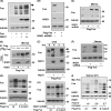
NQO1 inhibits c-Fos degradation by default. (A) HEK293 cells were transiently transfected with pCDNA-c-Fos with increasing amounts of pEFIRES-NQO1. Twenty-four hours after transfection, cells were lysed, and cell extracts were analyzed by Western blotting. An arrow marks the phosphorylated upshifted band. (B) MCF-7 cells were transfected with pCDNA-Flag-c-Fos alone or cotransfected with pSUPER empty vector or pSUPER-NQO1 expressing NQO1-specific shRNA. After 48 h, cell extracts were analyzed as described for panel A. (C) MCF-7 cells were treated as described for panel B, but 48 h after transfection, MG132 (10 μM) was added for 3 h. (D) HEK293 cells were transfected with the indicated plasmids. Twenty-four hours later, the cells were treated with 20 μM MG132 for 4 h, as indicated. Cells were labeled for 5 min with [35S]methionine, and samples were immunoprecipitated with anti-Flag antibody and subjected to autoradiography (IP) or immunoblotted (total). (E) HEK293 cells were transiently transfected with pCDNA-Flag-c-Fos without or with pEFIRES-NQO1. After 24 h, cells were treated with 20 μg/ml CHX for 3 h, and the extracts were analyzed as described for panel A. (F) MCF-7 cells were transfected with pCDNA encoding Flag-c-Fos together with pSUPER-NQO1shRNA or pSUPER vector. After 24 h, cells were starved for serum for 24 h and challenged with serum for 1 h, and the extracts were analyzed as described for panel A. (G) HEK293 cells were transiently transfected with pCDNA encoding either the wild type or the phosphorylation-deficient mutant (T232, T325, T331, S374A) (A mutant) without or with pEFIRES-NQO1. After 24 h, the samples were treated as described for panel A. (H) MCF-7 cells were transfected with pCDNA encoding either the wild type or the phosphorylation-deficient A mutant together with pSUPER-NQO1shRNA or pSUPER vector. After 48 h, the samples were treated as described for panel A. (I) To demonstrate that NQO1 protects c-Fos UID, we used A31N-ts20 cells stably overexpressing pEFIRES-NQO1 and the parental cell line stably transfected with the empty vector pEFIRES. WT c-Fos, a lysineless mutant (with no lysine residue even in the Myc tag) that is unable to undergo ubiquitination, and a phosphomimetic mutant (with S362D and S374D) (D mutant) were all accumulated by NQO1. Under the restrictive temperature of 39°C (24 h), only the UID takes place; hence, the accumulation of c-Fos by NQO1 provides strong evidence for NQO1 blocking c-Fos UID. The fold increase in Fos protein levels caused by NQO1 overexpression was quantified by the LAS-4000 software and is shown relative to the corresponding control sample.
FIG. 3.
FIG. 3.
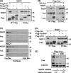
Role of c-Jun in the regulation of c-Fos degradation by default. (A) To examine the role of c-Jun in regulating c-Fos UID, MCF-7 cells were transfected with pCDNA-Flag-c-Fos alone or cotransfected with pCDNA-c-Jun. Twenty-four hours after transfection, cell extracts were analyzed by Western blotting. (B) To confirm that the upshifted band is the phosphorylated c-Fos, we used CIP. Cell extracts from the experiment described for panel A were incubated with CIP and analyzed by Western blotting. An arrow marks the phosphorylated upshifted band. (C) To demonstrate that c-Jun blocked c-Fos degradation by interfering with the UID process, A31N-ts20 cells were transfected with pCDNA-Flag-c-Fos without or with pCDNA-c-Jun at 39°C for 24 h, and the extracts were processed as described for panel A. (D) To examine the stability of c-Fos, we monitored the reduction level of the protein up to 6 h after blocking protein synthesis by CHX. HEK293 cells were transfected with pCDNA-Flag-c-Fos with or without pCDNA-c-Jun. After 24 h, the cell extracts were analyzed as described for panel A. (E) NQO1 is important for c-Jun to increase c-Fos accumulation. MCF-7 cells were transfected with pCDNA-Flag-c-Fos or pCDNA-c-Jun, alone or cotransfected with pSUPER-NQO1shRNA or pSUPER vector. After 48 h, cell extracts were analyzed as described for panel A. The level of c-Fos accumulation is much lower under NQO1 knockdown than in the presence of NQO1.
FIG. 4.
FIG. 4.
Role of the c-Fos leucine zipper in degradation by default. (A) To examine possible complex formation between NQO1 and c-Fos, HEK293 cells were transiently transfected with pCDNA-Flag-c-Fos, pEFIRES-Flag-p73α, or pEFIRES-Flag-p73β, together with pEFIRES-NQO1 (TOTAL). The p73α and p73β constructs were used as positive and negative controls, respectively. Immunoprecipitation of Flag-tagged proteins was performed with anti-Flag antibody-coupled beads (IP: Flag). The data clearly indicate that NQO1 is in association with c-Fos. (B) To examine whether NQO1 generates a tripartite complex with c-Fos and c-Jun, HEK293 cells were transiently transfected with pCDNA-Flag-c-Fos and pEFIRES-NQO1 without (−) or with (+) S3H HA-c-Jun (TOTAL). Immunoprecipitation of Flag-c-Fos was performed with anti-Flag beads (IP: Flag). The obtained data revealed that c-Jun interferes with NQO1-c-Fos complex formation. (C) HEK293 cells transfected with overexpressing c-Fos without or with NQO1 were lysed in RIPA buffer and loaded onto a 5 to 20% glycerol gradient. Afterward, ultracentrifugation fractions were collected and analyzed. The box with the dashed border shows the proteins shifted by NQO1 overexpression (lower panel) toward heavier complexes. (D) To assess the requirement of the c-Fos leucine zipper in NQO1 interaction, HEK293 cells were transiently transfected with pEFIRES-NQO1 with either pCDNA-Flag-c-Fos, the pCDNA-Flag-c-Fos VAV leucine zipper mutant, or pEFIRES-Flag-p73β (TOTAL). We performed immunoprecipitation of Flag-tagged proteins with anti-Flag beads (IP: Flag). The conclusion is that an intact leucine zipper is important for c-Fos-NQO1 complex formation. (E) To test the prediction that NQO1 will not support the c-Fos leucine zipper mutant, which does not associate with NQO1, MCF-7 cells were transfected with either pCDNA-Flag-c-Fos or the pCDNA-Flag-c-Fos VAV leucine zipper mutant alone or were cotransfected with pSUPER-NQO1shRNA or pSUPER vector. After 48 h, cell extracts were analyzed by Western blot analysis. Data show that the capacity of NQO1 to give rise to c-Fos accumulation is compromised once the leucine zipper is defective. The fold decrease in Fos protein levels caused by NQO1 knockdown was quantified by the LAS-4000 software and is shown relative to the corresponding control sample.
FIG. 5.
FIG. 5.
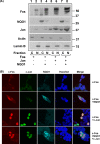
NQO1 and subcellular localization of c-Fos. (A) To determine protein subcellular localization, we first performed extract fractionation experiments. HEK293 cells were transfected with pCDNA-c-Fos alone or together with pEFIRES-NQO1 and pCDNA-c-Jun. Twenty-four hours after transfection, the cells were fractionated as described in Materials and Methods, and the equivalent amounts of the cytosolic (C) and soluble nuclear (N) fractions were submitted to immunoblot analysis. The localization of actin and β-lamin was analyzed to validate the fractionation procedure. The data show that nuclear accumulation of c-Fos is positively regulated by NQO1 and c-Jun. (B) Subcellular localization was visualized by confocal microscopy. HEK293 cells were transfected with pCDNA-Flag-c-Fos alone or together with pEFIRES-NQO1 and pCDNA-c-Jun. After 24 h, cells were fixed for immunofluorescence analysis with the indicated antibodies. Nuclear staining is indicated by using Hoechst 33342 stain.
FIG. 6.
FIG. 6.
NQO1 is required for efficient c-Fos serum induction. (A) To examine whether NQO1 overexpression affects the kinetics of endogenous c-Fos induction in response to serum induction, HCT116 p53−/− cells stably transfected with pEFIRES-NQO1 or with the empty vector were starved for serum for 24 h. The cells were then induced with serum, and the cell extracts were analyzed by Western blotting. c-Fos accumulation was more profound in HCT116 NQO1-overexpressing cells than in NQO1-deficient cells. (B) To estimate the kinetics of c-Fos serum induction under NQO1-deficient conditions, MCF-7 cells stably expressing NQO1 shRNAmir or control shRNAmir were starved for serum for 24 h. The cells were then induced with serum and analyzed as described for panel A. Under a low NQO1 level, c-Fos induction by serum was compromised, with a much lower rate of reaching a peak. (C) Serum induction of c-Fos was monitored under conditions devoid of ubiquitination and NQO1 knockdown. A31N-ts20 cells stably expressing NQO1 shRNA or the control shRNA were starved for serum for 24 h at 39°C. The cells were then incubated with serum for the indicated time points, lysed, and analyzed by Western blotting. Note the delay in appearance of the c-Fos peak from 1 to 2 h. (D) RT-PCR analysis on total RNA extracted from cells was done as in the experiment described for panel C. The level of c-Fos mRNA was found to be refractory to the level of NQO1.
FIG. 7.
FIG. 7.
NQO1-mediated c-Fos accumulation participates in a negative feedback loop. (A) Effect of NQO1 on the AP-1-responsive gene transcription. HEK293 cells were transfected with a luciferase reporter plasmid under the control of the collagenase promoter, together with pCDNA-c-Fos, pEFIRES-NQO1, or pEFIRES empty vector, as indicated. (B) MCF-7 cells were transfected with a luciferase reporter plasmid under the control of the collagenase promoter, together with pCDNA-c-Fos, pSUPER-NQO1shRNA, or empty pSUPER vector, as indicated. (C) NQO1 and c-Fos are interconnected by a feedback regulatory loop. H1299 cells were transfected with a luciferase reporter plasmid under the control of the nqo1 promoter, together with pCDNA-c-Fos, pEFIRES-NQO1, or pEGFP-MafF, as indicated. Luciferase activities were normalized by the transfection efficiency (shown as the means ± standard deviations), and the statistical significance (indicated by P values) was tested by the t test.
FIG. 8.
FIG. 8.
Steps in c-Fos expression; a simplified model to highlight our findings. According to our model, the newly synthesized c-Fos (arrow 1) is subjected to degradation by the 20S proteasome (arrow 2). NQO1 physically interacts with c-Fos to inhibit its degradation by default (arrow 3). Next, the AP-1 complex is formed (arrow 4), translocates to the nucleus, and is functional in DNA binding (arrow 5). At this stage, c-Fos degradation is regulated by ubiquitination and 26S-dependent proteasomal degradation (arrow 6).

Similar articles

Cited by

References

    1. Ahn, K. S., G. Sethi, A. K. Jain, A. K. Jaiswal, and B. B. Aggarwal. 2006. Genetic deletion of NAD(P)H:quinone oxidoreductase 1 abrogates activation of nuclear factor-kappaB, IkappaBalpha kinase, c-Jun N-terminal kinase, Akt, p38, and p44/42 mitogen-activated protein kinases and potentiates apoptosis. J. Biol. Chem. 281:19798-19808. - PubMed
    1. Asher, G., Z. Bercovich, P. Tsvetkov, Y. Shaul, and C. Kahana. 2005. 20S proteasomal degradation of ornithine decarboxylase is regulated by NQO1. Mol. Cell 17:645-655. - PubMed
    1. Asher, G., J. Lotem, B. Cohen, L. Sachs, and Y. Shaul. 2001. Regulation of p53 stability and p53-dependent apoptosis by NADH quinone oxidoreductase 1. Proc. Natl. Acad. Sci. U. S. A. 98:1188-1193. - PMC - PubMed
    1. Asher, G., J. Lotem, L. Sachs, C. Kahana, and Y. Shaul. 2002. Mdm-2 and ubiquitin-independent p53 proteasomal degradation regulated by NQO1. Proc. Natl. Acad. Sci. U. S. A. 99:13125-13130. - PMC - PubMed
    1. Asher, G., J. Lotem, P. Tsvetkov, V. Reiss, L. Sachs, and Y. Shaul. 2003. P53 hot-spot mutants are resistant to ubiquitin-independent degradation by increased binding to NAD(P)H:quinone oxidoreductase 1. Proc. Natl. Acad. Sci. U. S. A. 100:15065-15070. - PMC - PubMed

Publication types

MeSH terms