. 2010 Aug;54(8):3241-54.
doi: 10.1128/AAC.00364-10. Epub 2010 May 24.

Identification and characterization of small-molecule inhibitors of Yop translocation in Yersinia pseudotuberculosis

Affiliations

Identification and characterization of small-molecule inhibitors of Yop translocation in Yersinia pseudotuberculosis

Dana E Harmon et al. Antimicrob Agents Chemother. 2010 Aug.

Abstract

Type three secretion systems (TTSSs) are virulence factors found in many pathogenic Gram-negative species, including the family of pathogenic Yersinia spp. Yersinia pseudotuberculosis requires the translocation of a group of effector molecules, called Yops, to subvert the innate immune response and establish infection. Polarized transfer of Yops from bacteria to immune cells depends on several factors, including the presence of a functional TTSS, the successful attachment of Yersinia to the target cell, and translocon insertion into the target cell membrane. Here we employed a high-throughput screen to identify small molecules that block translocation of Yops into mammalian cells. We identified 6 compounds that inhibited translocation of effectors without affecting synthesis of TTSS components and secreted effectors, assembly of the TTSS, or secretion of effectors. One compound, C20, reduced adherence of Y. pseudotuberculosis to target cells. Additionally, the compounds caused leakage of Yops into the supernatant during infection and thus reduced polarized translocation. Furthermore, several molecules, namely, C20, C22, C24, C34, and C38, also inhibited ExoS-mediated cell rounding, suggesting that the compounds target factors that are conserved between Pseudomonas aeruginosa and Y. pseudotuberculosis. In summary, we have identified 6 compounds that specifically inhibit translocation of Yops into mammalian cells but not Yop synthesis or secretion.


Figures

FIG. 1.
FIG. 1.
HTS for small-molecule inhibitors of Yop translocation. (A) Fluorescence micrographs of HEp-2 cells loaded with CCF2-AM. (Left to right) Uninfected cells, HEp-2 cells infected with the WT E-TEM strain, and cells infected with the ΔyopB E-TEM strain. (B) Schematic of HTS. HEp-2 cells were loaded with CCF2-AM. Compounds were introduced by pin transfer, and Y. pseudotuberculosis (Yptb) was added at an MOI of 80:1. The compounds and Y. pseudotuberculosis were incubated together for 30 min before centrifugation to bring Y. pseudotuberculosis into contact with HEp-2 cells. After 60 min, the raw fluorescence values at 447 nm and 520 nm were determined on a plate reader.
FIG. 2.
FIG. 2.
YopE-mediated cell rounding was reduced after exposure of bacteria and HEP-2 cells to compounds. (A) Y. pseudotuberculosis was grown at 37°C in the presence of Ca2+ and compounds. HEp-2 cells were infected with WT IP2666 or IP2666 ΔyopB at an MOI of 10:1 in the presence of 60 μM compound (indicated in each panel). Images were taken at 45 min postinfection. (B) HEp-2 cells were cultured for 2 h in the presence of 60 μM compound in the absence of Y. pseudotuberculosis. Actin was visualized with FITC conjugated to phalloidin, and DAPI was used to visualize nuclei.
FIG. 3.
FIG. 3.
Compounds are not toxic to Y. pseudotuberculosis and have limited toxicity to HEp-2 cells. (A) Growth of Y. pseudotuberculosis was not inhibited by incubation with compounds. Y. pseudotuberculosis was grown in the presence of 60 μM compound or 0.3% DMSO for 7 h, and the OD600 was recorded each hour. ▪, DMSO; ▴, C7; ▾, C15; ⧫, C19; •, C20; □, C22; ▵, C24; ▿, C34; ⋄, C38. (B) LDH release from HEp-2 cells in the presence of 60 μM compound. The amount of LDH released into the supernatants was determined at 2 h and 24 h. The means and standard deviations from one representative experiment are plotted. (C) Structures of compounds that inhibited cell rounding of HEp-2 cells at a concentration of 60 μM and were not antibacterial or cytotoxic to HEp-2 cells.
FIG. 4.
FIG. 4.
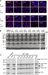
Localization and assembly of extracellular YscF and LcrV after exposure to compounds. (A) Y. pseudotuberculosis was grown in 2× YT supplemented with 5 mM CaCl2 at 37°C with a 60 μM concentration of the indicated compound or with DMSO for 1.5 h. Y. pseudotuberculosis was mounted and fixed on coverslips, labeled with anti-YscF or anti-LcrV antibody, and then visualized with Alexa Fluor 594-conjugated anti-rabbit antibody (red stain). Coverslips were counterstained with DAPI (blue stain). Images were pseudocolored and merged in MetaVue. (B) Y. pseudotuberculosis, grown in 3 mM CaCl2 with 60 μM compound or 0.3% DMSO, was treated with 1 mM BS3 or water. Y. pseudotuberculosis was solubilized, and Western blot analysis was performed with anti-YscF antibody. The asterisk shows YscF dimers, the arrowhead denotes YscF monomers, and the brace indicates high-molecular-weight YscF polymers. (C) Cultures of Y. pseudotuberculosis (Yptb) were grown in secretion medium (see Materials and Methods) with 60 μM compound. Yop secretion was detected by precipitation of cultured supernatants in 10% TCA. Proteins were separated by SDS-PAGE and stained with Coomassie blue to detect secreted Yops. The protein concentration was normalized to the OD, and equivalent amounts were loaded in each lane.
FIG. 5.
FIG. 5.
Translocation of YopE into HEp-2 cells reveals a translocation defect caused by compounds. Y. pseudotuberculosis (Yptb) was grown in 2× YT in the presence of 60 μM compound or 0.3% DMSO. Y. pseudotuberculosis was then used to infect HEp-2 cells at an MOI of 50:1 in the presence of 60 μM compound. After 1 h, the tissue culture supernatants were collected and the HEp-2 cells were lysed, whereupon the soluble fraction from HEp-2 cells and the insoluble fraction from the wells were collected. (A) HEp-2 cells were lysed, and cytosol fractions were separated by SDS-PAGE and probed with antiserum to YopE. The percent translocation was determined by measuring the amount of YopE in the soluble fraction and normalizing it to the amount of S2 protein in the insoluble fraction. This ratio was then normalized to the amount of actin as a loading control. The amount of YopE translocated by WT Y. pseudotuberculosis in 0.3% DMSO was set to 100% translocation. (B) The insoluble fraction was separated by SDS-PAGE, and Western blot analysis was performed with anti-YopE and anti-S2 antibodies. The levels of YopE were normalized to the level of S2 (percent YopE synthesis). The amount of YopE in the DMSO-treated sample was set to 100%. (C) Tissue culture supernatants were collected, and bacteria were removed by centrifugation. Anti-YopE antibody was used to immunoprecipitate YopE protein. The proteins collected were separated by SDS-PAGE and subjected to Western blot analysis with antiserum to YopE. The amount of leaked YopE was determined by normalizing YopE protein levels from the immunoprecipitated fraction to S2 levels from the insoluble fraction. The ratios for compound-containing wells to DMSO controls were graphed. For all panels, the means and standard deviations for three independent experiments are plotted. Representative Western blots are also shown. *, P ≤ 0.05; **, P ≤ 0.06.
FIG. 6.
FIG. 6.
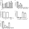
Adherence of Y. pseudotuberculosis to HEp-2 cells is reduced by C20. (A) Adherence of Y. pseudotuberculosis to HEp-2 cells in the presence of compounds was determined by ELISA. WT Y. pseudotuberculosis was incubated with 60 μM compound. HEp-2 cells were infected in the presence of compounds with Y. pseudotuberculosis at an MOI of 10:1 for 30 min at 37°C. HEp-2 cells were washed to remove any unbound Y. pseudotuberculosis, fixed with 4% paraformaldehdye, and then probed with antisera to Yersinia. Anti-rabbit-HRP was used to detect anti-Yersinia antibody. The HRP activity was visualized with the TMB ELISA reagent, and the DMSO control was set to 100% adherence. The means and standard deviations for one representative experiment are plotted. (B) WT Y. pseudotuberculosis or adherence mutants were grown in 2× YT supplemented with 5 mM Ca2+ with C20 or 0.3% DMSO and were used to infect HEp-2 cells. The percent adherence was determined by ELISA as described above. The percent adherence of WT Y. pseudotuberculosis grown in 0.3% DMSO was set to 100%. The means and standard deviations for one representative experiment are plotted. (C) Overnight cultures of Y. pseudotuberculosis were inoculated into RPMI and allowed to incubate statically at 37°C for 3 h in the presence or absence of C20. Autoagglutination was measured as described in Materials and Methods. The percent autoagglutination of WT IP2666 in 0.3% DMSO was set to 100%. The means and standard deviations for one representative experiment are shown. (D) E. coli carrying pDS132 or pDS132-yadA was cultured in 60 μM C20 or in 0.3% DMSO and was used to infect HEp-2 cells in the presence of C20 or DMSO. Adherence of E. coli to HEp-2 cells was determined by ELISA as described for panel A, using antiserum to the E. coli phage λ receptor, LamB. The means and standard deviations for one representative experiment are plotted. (E) Hemolysis of SRBCs by Y. pseudotuberculosis adherence mutants or WT Y. pseudotuberculosis grown in the presence of 60 μM C20. Y. pseudotuberculosis was grown in secretion medium in the presence of either 0.3% DMSO or C20 and was used to infect SRBCs at an MOI of 1:1. The percent hemolysis was measured by the amount of hemoglobin released from the SRBCs (see Materials and Methods). Hemolysis by WT Y. pseudotuberculosis in 0.3% DMSO was set to 100%. The means and standard deviations for one representative experiment are shown.
FIG. 7.
FIG. 7.
Pseudomonas aeruginosa ExoS-dependent cell rounding was blocked by C20, C22, C24, C34, and C38. Cultures of WT P. aeruginosa (Pa388) or a translocation-defective mutant (Pa388 pscC) were grown at 37°C in the presence of 60 μM compound or 0.3% DMSO. The cultures were used to infect HEp-2 cells at an MOI of 10:1 in the presence of 60 μM compound. The infection was allowed to proceed for 90 min at 37°C before imaging. The experiment was repeated twice, and representative micrographs are shown.
FIG. 8.
FIG. 8.
Summary of results for compounds identified in the screen for small-molecule inhibitors of Yop translocation. Yptb, Y. pseudotuberculosis; Pa, P. aeruginosa.

References

    1. Achtman, M., K. Zurth, G. Morelli, G. Torrea, A. Guiyoule, and E. Carniel. 1999. Yersinia pestis, the cause of plague, is a recently emerged clone of Yersinia pseudotuberculosis. Proc. Natl. Acad. Sci. U. S. A. 96:14043-14048. - PMC - PubMed
    1. Aiello, A. E., G. F. Murray, V. Perez, R. M. Coulborn, B. M. Davis, M. Uddin, D. K. Shay, S. H. Waterman, and A. S. Monto. 2010. Mask use, hand hygiene, and seasonal influenza-like illness among young adults: a randomized intervention trial. J. Infect. Dis. 201:491-498. - PubMed
    1. Aiello, D., J. D. Williams, H. Majgier-Baranowska, I. Patel, N. P. Peet, J. Huang, S. Lory, T. L. Bowlin, and D. T. Moir. 2010. Discovery and characterization of inhibitors of Pseudomonas aeruginosa type III secretion. Antimicrob. Agents Chemother. 54:1988-1999. - PMC - PubMed
    1. Aili, M., E. L. Isaksson, S. E. Carlsson, H. Wolf-Watz, R. Rosqvist, and M. S. Francis. 2008. Regulation of Yersinia Yop-effector delivery by translocated YopE. Int. J. Med. Microbiol. 298:183-192. - PubMed
    1. Bailey, L., A. Gylfe, C. Sundin, S. Muschiol, M. Elofsson, P. Nordstrom, B. Henriques-Normark, R. Lugert, A. Waldenstrom, H. Wolf-Watz, and S. Bergstrom. 2007. Small molecule inhibitors of type III secretion in Yersinia block the Chlamydia pneumoniae infection cycle. FEBS Lett. 581:587-595. - PubMed

Publication types

MeSH terms