. 2010 May;10(2):159-73.
doi: 10.3758/CABN.10.2.159.

Resisting emotional interference: brain regions facilitating working memory performance during negative distraction

Affiliations

Resisting emotional interference: brain regions facilitating working memory performance during negative distraction

Alan Anticevic et al. Cogn Affect Behav Neurosci. 2010 May.

Abstract

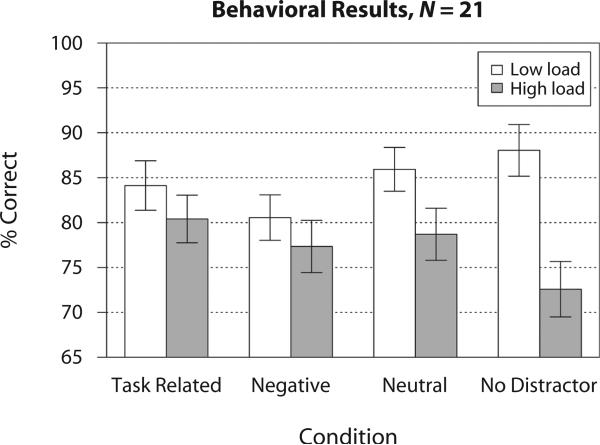
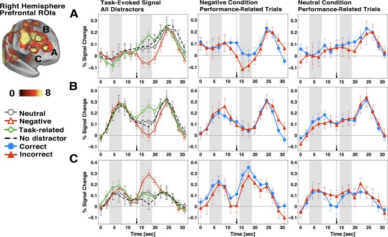
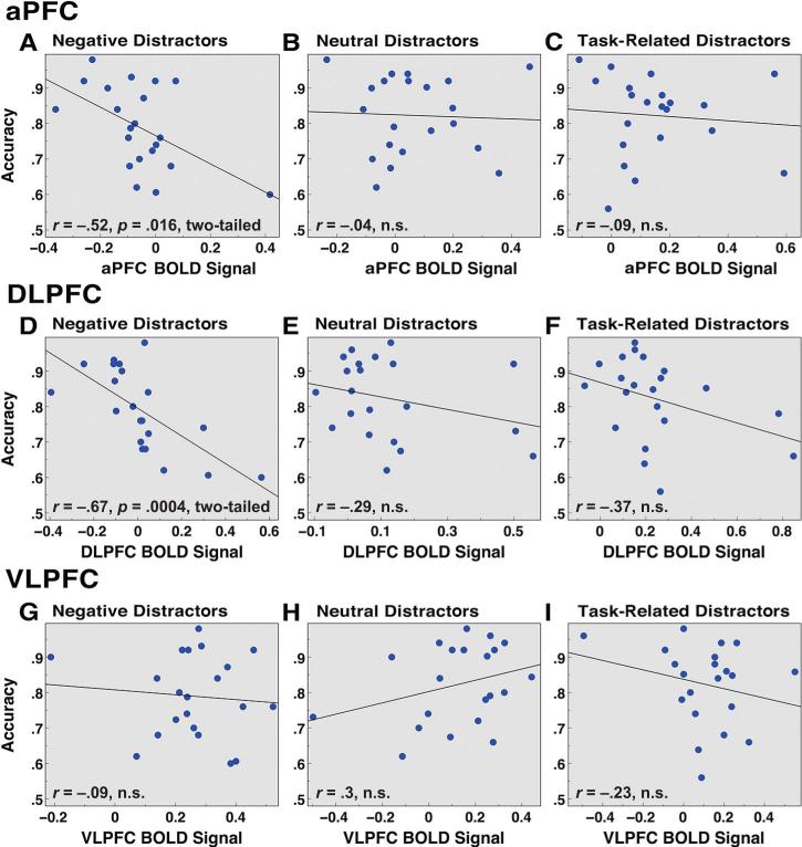
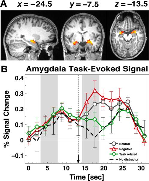
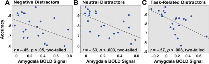
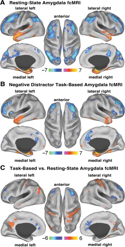
Survival-relevant information has privileged access to our awareness even during active cognitive engagement. Previous work has demonstrated that during working memory (WM) negative emotional distraction disrupts activation in the lateral prefrontal regions while also engaging the amygdala. Here, using slow event-related fMRI, we replicate and extend previous work examining the effect of negative emotional distraction on WM: (1) We demonstrate that prefrontal regions showed activation differences between correct and incorrect trials during negative, but not neutral, distraction. Specifically, frontopolar prefrontal cortex showed more deactivation for incorrect trials faced with negative distraction, whereas ventrolateral prefrontal regions showed less activation; (2) individual differences in amygdala activity predicted WM performance during negative as well as neutral distraction, such that lower activity predicted better performance; and (3) amygdala showed negative correlations with prefrontal and parietal cortical regions during resting state. However, during negative distraction, amygdala signals were more negatively correlated with prefrontal cortical regions than was found for resting state and neutral distraction. These results provide further evidence for an inverse relationship between dorsal prefrontal cortical regions and the amygdala when processing aversive stimuli competes with ongoing cognitive operations, and further support the importance of the prefrontal cortex in resisting emotional interference. Supplemental materials associated with this article may be downloaded from http://cabn.psychonomic-journals.org/content/supplemental.


Figures

Figure 1
Figure 1
Task design. The overall layout of the task is shown along with different components and their onsets marked along the timeline. each box represents a trial component with the duration marked below. First, subjects were presented with a set of complex geometric shapes, which they were instructed to memorize, followed by a delay. Next, during the middle phase of the trial subjects saw either (1) an emotionally negative distractor; (2) a task-related geometric shape distractor of a different color distinguishing it from the probe; (3) a neutral distractor; or (4) no distraction. This was followed by another delay. Finally, subjects were shown a probe and indicated via a button response whether it was part of the memorized set or not.
Figure 2
Figure 2
Behavioral results. Mean accuracy (expressed as percent correct) is shown for task-related, negative, neutral, and nodistractor conditions across two load levels. Error bars represent ±1 standard error of the mean.
Figure 3
Figure 3
Task-evoked and performance-related time courses for right lateral prefrontal cortical foci. event-related time courses are shown for right (A) anterior prefrontal cortex; (B) dorsolateral prefrontal cortex; and (C) ventrolateral prefrontal cortex. The far left set of graphs shows the task-evoked signals for neutral (circles), negative (triangles), task-related (diamonds), and no-distractor (dashed lines) conditions across all three RoIs. The middle panel shows performance-related time courses for the negative condition. The far right panel shows performance-related time courses for the neutral condition. Correct and incorrect time courses are shown with circles and triangles, respectively. Distractor onset is marked with a dotted vertical line ending in an arrow.
Figure 4
Figure 4
Individual differences in working memory (WM) performance as a function of prefrontal signal. Average WM performance (proportion correct) is shown as a function of average signal in PFC RoIs. (A–C) Frontopolar prefrontal cortex (aPFC) RoI is shown for negative (r = –.52, p = .016, two-tailed), neutral (r = –.04, n.s.), and task-related (r = –.09, n.s.) distractor conditions; (D–F) DlPFC RoI is shown for negative (r = –.67, p = .0004, two-tailed), neutral (r = –.29, n.s.), and task-related (r = –.37, n.s.) distractor conditions; (G–I) VlPFC RoI is shown for negative (r = –.09, n.s.), neutral (r = .3, n.s.), and task-related (r = –.23, n.s.) distractor conditions. Results are collapsed across low and high WM load given the same pattern at both loads. PFC BolD signal was averaged across both correct and incorrect trials.
Figure 5
Figure 5
Bilateral amygdala signal. (A) Bilateral amygdala activation maps and (B) bilateral amygdala time courses are shown for neutral (circles), negative (triangles), task-related (diamonds), and no-distractor (dashed lines) conditions. Distractor onset is marked with a dotted vertical line ending in an arrow.
Figure 6
Figure 6
Individual differences in working memory (WM) performance as a function of amygdala signal. Average WM performance (proportion correct) is shown as a function of average bilateral amygdala signal for (A) negative (r = –.45, p < .05, two-tailed); (B) neutral (r = –.63, p < .003, two-tailed); and (C) task-related (r = –.57, p < .008, two-tailed) distractor conditions. Results are collapsed across low and high WM load given the same pattern at both loads. Amygdala BolD signal was averaged across both correct and incorrect trials.
Figure 7
Figure 7
Amygdala resting-state and task-based functional connectivity. All maps are shown using Z statistics and visualized using the PAlS atlas (Van essen, 2005). Bilateral amygdala fcMRI is shown (A) during resting state and (B) during WM faced with negative distraction. Brighter colors mark regions showing either more positive or more negative fcMRI with amygdala. The online version of this article shows positive and negative fcMRI with the amygdala in orange-yellow and blue colors, respectively. Both resting-state and task-based fcMRI maps show results corrected at whole-brain p < .05. (C) We also show results of an independent samples t test comparing resting-state and task-based amygdala fcMRI. here we show foci using a Z > 2.5 threshold demonstrating that even with a lower cutoff more negative fcMRI for task versus resting state is centered mainly around the pre-frontal nodes network and not elsewhere. The same foci are also shown using a whole-brain p < .05 correction in Supplemental Figure S5.

References

    1. Aaron AR, Robbins TW, Poldrack RA. Inhibition and the right inferior frontal cortex. Trends in Cognitive Sciences. 2004;8:170–177. - PubMed
    1. Attneave F, Arnoult MD. The quantitative study of shape and pattern perception. Psychological Bulletin. 1956;53:452–471. - PubMed
    1. Avenanti A, Bueti D, Galati G, Aglioti SM. Transcranial magnetic stimulation highlights the sensorimotor side of empathy for pain. Nature Neuroscience. 2005;8:955–960. - PubMed
    1. Avenanti A, Minio-Paluello I, Bufalari I, Aglioti SM. Stimulus-driven modulation of motor-evoked potentials during observation of others’ pain. NeuroImage. 2006;32:316–324. - PubMed
    1. Baddeley AD, Hitch GJ. Developments in the concept of working memory. Neuropsychology. 1994;8:485–493.

Publication types

LinkOut - more resources