. 2010 May 13;5(5):e10626.
doi: 10.1371/journal.pone.0010626.

Effective melanoma immunotherapy in mice by the skin-depigmenting agent monobenzone and the adjuvants imiquimod and CpG

Affiliations

Effective melanoma immunotherapy in mice by the skin-depigmenting agent monobenzone and the adjuvants imiquimod and CpG

Jasper G van den Boorn et al. PLoS One. .

Abstract

Background: Presently melanoma still lacks adequate treatment options for metastatic disease. While melanoma is exceptionally challenging to standard regimens, it is suited for treatment with immunotherapy based on its immunogenicity. Since treatment-related skin depigmentation is considered a favourable prognostic sign during melanoma intervention, we here aimed at the reverse approach of directly inducing vitiligo as a shortcut to effective anti-melanoma immunity.

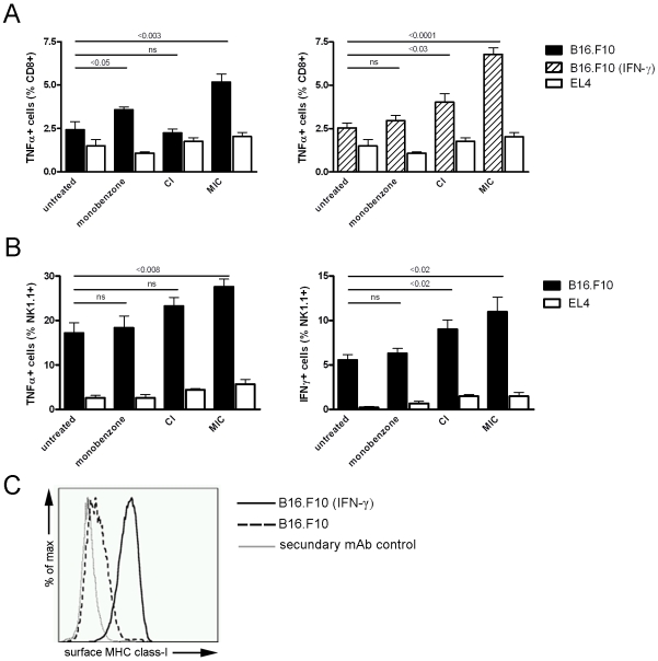
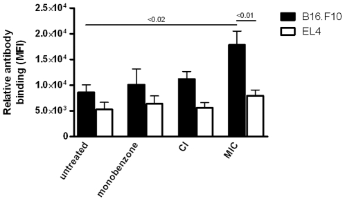
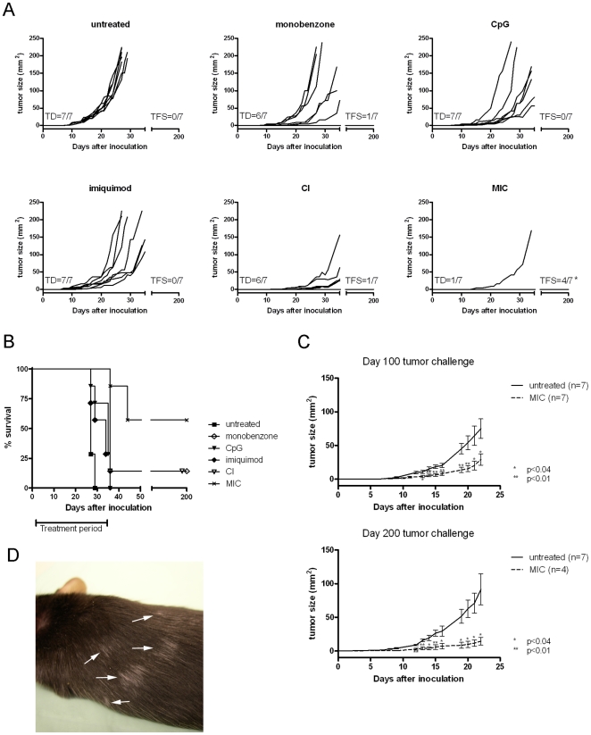
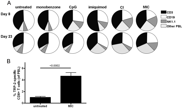
Methodology and principal findings: We developed an effective and simple to use form of immunotherapy by combining the topical skin-bleaching agent monobenzone with immune-stimulatory imiquimod cream and cytosine-guanine oligodeoxynucleotides (CpG) injections (MIC therapy). This powerful new approach promptly induced a melanoma antigen-specific immune response, which abolished subcutaneous B16.F10 melanoma growth in up to 85% of C57BL/6 mice. Importantly, this regimen induced over 100 days of tumor-free survival in up to 60% of the mice, and forcefully suppressed tumor growth upon re-challenge either 65- or 165 days after MIC treatment cessation.

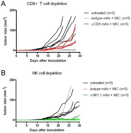
Conclusions: MIC therapy is effective in eradicating melanoma, by vigilantly incorporating NK-, B- and T cells in its therapeutic effect. Based on these results, the MIC regimen presents a high-yield, low-cost and simple therapy, readily applicable in the clinic.


Conflict of interest statement

Competing Interests: The authors have declared that no competing interests exist.

Figures

Figure 1
Figure 1. MIC treatment of subcutaneous B16.F10 melanoma induced melanoma-reactive CD8+ T cells and -NK cells in vivo.
Splenocytes were tested for their ex vivo activation upon co-culture with B16.F10 melanoma or EL4 thymoma control cells (n = 5 mice per group). A, Left panel: CD8+ T cells from monobenzone- and MIC-treated mice showed significant TNF-α production upon co-culture with melanoma cells (black bars; p<0.05 and p<0.003 respectively). In contrast, CD8+ T cells from CI-treated mice did not display significant TNF-α production upon melanoma cell co-culture (non significant difference: ns). Right panel: To identify CD8+ T cell activation upon co-culture with immunogenic melanoma cells with high MHC class-I expression, co-cultures with IFN-γ primed B16.F10 cells were included (dashed bars). Under these conditions, CI-treated mice showed significant TNF-α production as compared to untreated mice (p<0.03). The MIC-treated mice showed even more TNF-α production as in the non-IFN-γ-primed stimulation shown in the left panel. Monobenzone-treated and untreated mice did not display this increased T cell activation upon splenocyte co-culture with IFN-γ primed melanoma cells. T cell activation upon splenocyte co-culture with syngeneic EL4 thymoma control cells showed comparable background levels in all groups (white bars). B, Left panel: Only NK cells from MIC-treated mice showed significantly increased TNF-α production upon co-culture with melanoma cells (p<0.008). Right panel: Elevated production of IFN-γ was found in NK cells from CI- and MIC-treated mice in response to co-culture with melanoma cells (p<0.02 and p<0.02 respectively). TNF-α and IFN-γ production by NK cells was comparable in all groups upon co-culture with EL4 control cells. For the statistical analysis of the in vivo tumor growth kinetics of the treatments depicted in this figure, see table 1 (“Exp. 2”). C, B16.F10 melanoma cells upregulate their surface MHC class-I expression upon IFN-γ exposure. While IFN-γ-unexposed melanoma cells express very low levels of surface MHC class-I (dashed line), priming of these cells with 1000 U/ml IFN-γ restores their surface expression of MHC class-I (black line). Control incubations of IFN-γ-primed melanoma cells with only the IgG2a-detecting secondary antibody were negative (grey line).
Figure 2
Figure 2. A melanoma-reactive serum IgG response was found in MIC-treated mice.
Upon sacrifice peripheral blood serum was obtained from treated mice, and serum antibody binding to B16.F10 melanoma cells was analyzed using flow cytometry (n = 5 mice per group). EL4 syngeneic thymoma cells were used as a control to verify the melanoma-specificity of the antibody binding, and showed comparable serum IgG binding levels in all groups (p>0.05, one-way ANOVA). A significant level of IgG antibody binding to the melanoma cells above the EL4 background level was found in the MIC-treated mice (p<0.01, paired t-test). Furthermore, only the MIC-treated mice showed melanoma-reactive IgG levels significantly above those found in untreated mice (p<0.02, unpaired t-test). IgA controls were negative, and IgM antibodies showed no significant binding levels (data not shown). Data is representative of three independent in vivo experiments.
Figure 3
Figure 3. Growth of subcutaneous melanoma is inhibited by MIC therapy.
Mice (n = 7 per group) were treated with monobenzone, CpG, imiquimod, CI- or the MIC-regimen. Tumor growth and animal survival were monitored for 200 days. Each graph line depicts an individual tumor growth curve. A, Upper left panel: Untreated mice all show tumor development (TD) around day 10, and none of the mice experienced a 200-day tumor-free survival (TFS). In contrast, 6 out of 7 MIC-treated mice remained tumor-free during treatment, and only one mouse showed delayed TD during the treatment (lower right panel). *:10 days following treatment cessation at day 35, two additional mice developed a melanoma and these animals were directly sacrificed for analysis without monitoring tumor growth kinetics. Eventually, 4 out of 7 MIC-treated mice showed 200-day TFS, since 2 of the mice developed a melanoma 10 days following treatment cessation at day 35. The individual treatment components monobenzone, CpG or imiquimod all mediated a certain degree of tumor-growth delay. Interestingly, CpG and imiquimod clearly work synergistically in the CI regimen. Depicted tumor growth kinetics are representative of 4 independent in vivo experiments. For the statistical analysis of the in vivo tumor growth kinetics of the treatments depicted in this figure, see table 1 (“Exp. 4”). B, Kaplan-Meier survival curve for the different treatment groups depicted under A, showing 57% 200-day survival for MIC-treated mice against 14% long-term survival for monobenzone- or CI treated mice. Mice left untreated or receiving one of the individual treatment components, no TFS was found. These animals were sacrificed around day 25–35 due to maximally allowed tumor burden. C, The MIC therapy induced an effective immunological memory response. Upper panel: At day 100 (day 65 after treatment cessation) surviving mice were challenged with a melanoma tumor-inoculation, and tumor growth was monitored without further treatment. Untreated naive control mice showed rapid tumor development (black line, n = 7). Mice treated previously with MIC therapy showed significant tumor growth delay (dashed line, n = 7). Lower panel: Mice challenged at day 200 (day 165 after treatment cessation) show protective immunity to a comparable degree to that found in the day 100 tumor challenge shown in the upper panel. Untreated control mice showed normal tumor development (black line, n = 7) while mice treated previously with the MIC therapy display significant tumor growth retardation (dashed line, n = 4). Depicted graphs represent two independent tumor challenge experiments (follow-up on Exp. 3 & 4 in table 1). Statistical analysis using unpaired t-test comparing MIC-treated mice with untreated animals on designated time points (*: p<0.04, **:p<0.01). D, MIC-treated, long-term surviving mice occasionally develop progressively depigmenting patches of fur in a vitiligo-like pattern, distant from the initial monobenzone application site (arrows). This effect occurs in approximately 50% of the long-term surviving animals.
Figure 4
Figure 4. MIC-treated mice show a sustained NK cell expansion and circulating melanocyte antigen-specific CD8+ T cells.
A, Peripheral blood was collected from the tailvein of treated mice on day 8 and 23 of treatment, and average ratios between T cells (CD3+, black sections), B cells (CD19+, white sections), NK cells (NK1.1+, CD3-, grey sections) and other peripheral blood leukocytes (PBL; dashed sections) were determined for the PBL (n = 7 mice per group). Interestingly, MIC-treated mice showed a significant NK cell expansion on both day 8 and 23 (see table 2, “Exp. 4”). This expansion of NK cells was also found in CpG-, imiquimod- and CI-treated mice, although for these animals this reaction was only found on day 8. Monobenzone alone did not influence PBL ratios on either time point, comparable to untreated mice. Depicted data is representative of three independent in vivo mouse experiments. For the statistical analysis of the in vivo differences in NK cell counts in these experiments see table 2. B, Peripheral blood CD8+ T cells were tested for binding to H2-Kb/TRP-2180-188-tetramers at day 120 following tumor inoculation (day 85 after treatment cessation, n = 4). TRP-2 represents one of the immunodominant epitopes of B16.F10 melanoma. Long-term surviving, MIC-treated mice showed a significant population of TRP-2-specific CD8+ T cells circulating in their peripheral blood at day 120, as compared to untreated mice 10 days after tumor inoculation (n = 7). Binding to control H2-Kb/OVA257-264-tetramer by the tested PBL was negative (data not shown).
Figure 5
Figure 5. The therapeutic effect of MIC therapy is abrogated by NK cell- or CD8+ T cell depletion.
Each graph line represents an individual tumor growth curve. A, Mice depleted of CD8+ T cells prior to tumor inoculation and throughout the MIC therapy (red lines, n = 5) all developed a tumor. In these MIC-treated mice tumors grew out significantly faster than in the isotype-control mAb-treated mice (grey lines; p<0.02, n = 5), who displayed tumor growth kinetics similar to MIC-treated animals in previous experiments. Nonetheless, tumor outgrowth in CD8+ T cell depleted mice was still significantly slower than in untreated mice (black lines; p<0.02, n = 5). The tumor size of CD8+ T cell depleted animals was statistically compared with isotype-control mAb-treated animals on day 28, and with untreated animals on day 22. B, Mice depleted of NK cells prior to tumor inoculation and throughout MIC therapy (green lines, n = 5) showed tumor establishment in 80% of mice. Tumors in these mice grew out very slowly or remained of equal small size throughout the experiment. In contrast, untreated mice showed rapid tumor development (black lines, n = 5). Mice treated with isotype-control mAb and MIC therapy (grey lines, n = 5) showed tumor growth similar to mice treated with MIC therapy in previous experiments.

References

    1. Smith JL, Jr, Stehlin JS., Jr Spontaneous regression of primary malignant melanomas with regional metastases. Cancer. 1965;18:1399–1415. - PubMed
    1. Chen KG, Leapman RD, Zhang G, Lai B, Valencia JC, et al. Influence of melanosome dynamics on melanoma drug sensitivity. J Natl Cancer Inst. 2009;101:1259–1271. djp259 [pii];10.1093/jnci/djp259 [doi] - PMC - PubMed
    1. Bart RS, Porzio NR, Kopf AW, Vilcek JT, Cheng EH, et al. Inhibition of growth of B16 murine malignant melanoma by exogenous interferon. Cancer Res. 1980;40:614–619. - PubMed
    1. Kirkwood JM, Strawderman MH, Ernstoff MS, Smith TJ, Borden EC, et al. Interferon alfa-2b adjuvant therapy of high-risk resected cutaneous melanoma: the Eastern Cooperative Oncology Group Trial EST 1684. J Clin Oncol. 1996;14:7–17. - PubMed
    1. Rosenberg SA, Mule JJ, Spiess PJ, Reichert CM, Schwarz SL. Regression of established pulmonary metastases and subcutaneous tumor mediated by the systemic administration of high-dose recombinant interleukin 2. J Exp Med. 1985;161:1169–1188. - PMC - PubMed

Publication types

MeSH terms