. 2010 May 13;5(5):e10627.
doi: 10.1371/journal.pone.0010627.

Superoxide dismutase 1 and tgSOD1 mouse spinal cord seed fibrils, suggesting a propagative cell death mechanism in amyotrophic lateral sclerosis

Affiliations

Superoxide dismutase 1 and tgSOD1 mouse spinal cord seed fibrils, suggesting a propagative cell death mechanism in amyotrophic lateral sclerosis

Ruth Chia et al. PLoS One. .

Abstract

Background: Amyotrophic lateral sclerosis (ALS) is a neurodegenerative disease that specifically affects motor neurons and leads to a progressive and ultimately fatal loss of function, resulting in death typically within 3 to 5 years of diagnosis. The disease starts with a focal centre of weakness, such as one limb, and appears to spread to other parts of the body. Mutations in superoxide dismutase 1 (SOD1) are known to cause disease and it is generally accepted they lead to pathology not by loss of enzymatic activity but by gain of some unknown toxic function(s). Although different mutations lead to varying tendencies of SOD1 to aggregate, we suggest abnormal proteins share a common misfolding pathway that leads to the formation of amyloid fibrils.

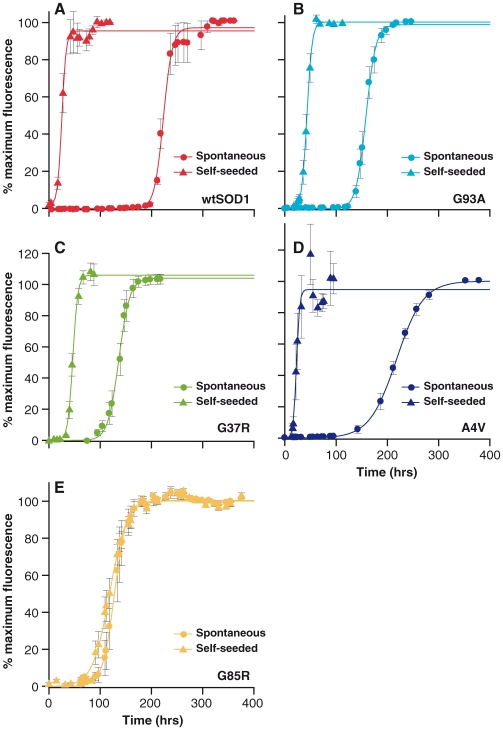
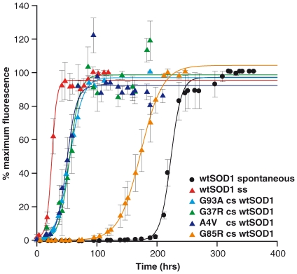
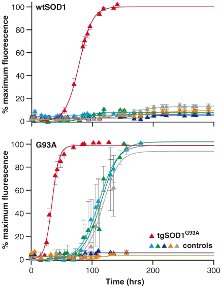
Methodology/principal findings: Here we demonstrate that misfolding of superoxide dismutase 1 leads to the formation of amyloid fibrils associated with seeding activity, which can accelerate the formation of new fibrils in an autocatalytic cascade. The time limiting event is nucleation to form a stable protein "seed" before a rapid linear polymerisation results in amyloid fibrils analogous to other protein misfolding disorders. This phenomenon was not confined to fibrils of recombinant protein as here we show, for the first time, that spinal cord homogenates obtained from a transgenic mouse model that overexpresses mutant human superoxide dismutase 1 (the TgSOD1(G93A) mouse) also contain amyloid seeds that accelerate the formation of new fibrils in both wildtype and mutant SOD1 protein in vitro.

Conclusions/significance: These findings provide new insights into ALS disease mechanism and in particular a mechanism that could account for the spread of pathology throughout the nervous system. This model of disease spread, which has analogies to other protein misfolding disorders such as prion disease, also suggests it may be possible to design assays for therapeutics that can inhibit fibril propagation and hence, possibly, disease progression.


Conflict of interest statement

Competing Interests: The authors have declared that no competing interests exist.

Figures

Figure 1
Figure 1. Conditions screened for SOD1 fibrillisation.
SOD1 variants with higher stability were able to form fibrils over a wide range of conditions (i.e. number of combinations of pH and denaturant strength) characterized by variation in lag times (wtSOD red square = 10 combinations; G93A light blue square = 6 combinations; G37R green square = 5 combinations; A4V dark blue square = 2 combinations; G85R yellow square = 2 combinations). For the more stable proteins, when incubated in increasing denaturant strength, lag time for fibrils to form is shortened (data not shown).
Figure 2
Figure 2. Spontaneous and self-seeded fibrillization of SOD1.
Spontaneous and self-seeded reactions for (A) wtSOD1, (B) G93A, (C) G37R, (D) A4V, and (E) G85R. All SOD1 variants, except G85R were incubated at 37°C, at pH4.0 in mild denaturing condition (0.5M GdnHCl). Fibrillization of G85R was performed at pH5.0 with no denaturant. Fibril formation is reported as % maximum ThT relative fluorescence units (RFU) shown as a function of time (hrs). Seeded reactions were spiked with 1% preformed fibrils (v/v). Data shown are average values from 6–10 replicates (± SEM) pooled from 2–3 independent assays. Seeded fibrillization reactions consistently shortened the lag times by an average of 82% (except for G85R) compared to spontaneous reactions under similar conditions.
Figure 3
Figure 3. Cross-seeding (cs) fibrillization reactions with preformed fibrils of SOD1 variants into wtSOD1.
Formation of fibrils are reported as % maximum ThT relative fluorescence units (RFU) shown as a function of time. Seeded reactions were spiked with 1% preformed fibrils (v/v). Data shown are average values from 6–10 replicates (± SEM) pooled from 2–3 independent assays. Self-seeding of wtSOD1 fibrils into recombinant wtSOD1 (wtSOD1 ss) gave the greatest reduction in lag time with cross-seeding of G93A, G37R, and A4V into wtSOD1 shortening the lag time from 206 hours to between 33 and 38 hours (lag time shortened by approximately 83%). G85R was also capable of reducing the lag time for wtSOD1 fibrillisation but less significantly (33% reduction in lag time).
Figure 4
Figure 4. Fibrillogenic seeding with affected tissue homogenates.
wtSOD1 (top graph) and G93A (bottom graph) proteins were seeded with spinal cord homogenates from 120 days old tgSOD1G93A (red triangle), tgSOD1wtSOD1 (light blue triangle) and control non-transgenic littermates of the tgSOD1G93A (green triangle) mice. Seeded reactions spiked with 1% (v/v) tgSOD1G93A spinal cord homogenates into wt SOD1 and G93A proteins shortened the lag times to approximately 56 hrs and 23hrs respectively, comparable to seeded reactions with pre-formed recombinant G93A fibrils to wt SOD1 and to self-protein (G93A) (refer to Table 1). The lag times of seeded reactions with tgSOD1wtSOD1 and non-transgenic control spinal cord homogenates into G93A protein were 1.5–3.5 fold longer than the lag times of seeded reactions by tgSOD1G93A spinal cord homogenates into G93A protein. Seeding with tgSOD1wtSOD1 and non-transgenic control spinal cord homogenates into wtSOD1 protein did not give rise to any ThT fluorescence change, indicating an absence of fibril formation. Additional spinal cord homogenate controls were used to test for seeding specificity as described in the text: spinal cord homogenates were prepared from clinical endstage Tg20 mice infected with the RML strain of prions (dark blue triangle), huntingtin transgenic mice (N171-82Q) (grey triangle), and brain homogenate prepared derived from an FTD3 patient (yellow triangle).Data shown are average values from 4–10 replicates (± SEM) pooled from 2–3 independent assays.
Figure 5
Figure 5. Representative electron micrographs of SOD1 fibrils from spontaneous and seeded reactions (self-seeded and cross-seeded with pre-formed fibril/oligomer or with spinal cord tissue homogenate).
Samples from fibrillization reactions of (A) A4V spontaneous, (B) A4V self-seeded, (C) A4V cross-seeded samples into wtSOD1 protein, (D) G37R spontaneous, (E) G85R spontaneous, (F) G85R self-seeded, (G) G93A spontaneous, (H–I) tgSOD1G93A spinal cord homogenate seeded into G93A protein. All spontaneous, self- and cross-seeded reactions for all SOD1 variants were carried out at pH4.0 with 0.5M GdnHCl, except for G85R which was carried out at pH5.0 without any denaturant. Shorter fibrillar fragments are indicated by yellow arrows, which were observed in all samples. Green arrows point to longer and continuous fibrillar threads, whereas black arrows indicate thinner and less regular SOD1 species (possibly shorter oligomers). Blue arrows in (E) point to a less structured morphology of G85R ThT-positive species (most appear as spherical aggregates but some have a toroidal appearance) a morphology which was also rarely observed in other SOD1 fibrillar samples. Red arrows in (H) and (I) indicate flexion points of what appears to be 2–3 protofilaments together to form a thicker fibrillar thread or fragment. All SOD1 fibril samples which were qualitatively characterized under EM showed a common property of increased propensity to clump together or to aggregate, regardless of the morphology of the ThT-positive species. Scale bar = 400nm.
Figure 6
Figure 6. The effect of protein stability on the lag time of SOD1 fibrillization for spontaneous and self-seeded reactions.
The plot shows the correlation of SOD1 stability, in the metallated and intra-subunit intact form, (expressed as melting temperature, Tm (°C)) to lag time (hrs). Destabilisation of SOD1 correlated with shorter lag times for fibrillisation in spontaneous reactions, whereas reactions initiated with pre-formed fibrils had lag times that were independent of the original native fold stability. (Tm data for G85R not available, thus it was not possible to include the G85R fibrillization lag time and propagation rate in the graphs.)

References

    1. Dion PA, Daoud H, Rouleau GA. Genetics of motor neuron disorders: new insights into pathogenic mechanisms. Nat Rev Genet. 2009;10:769–782. - PubMed
    1. Ince PG, Lowe J, Shaw PJ. Amyotrophic lateral sclerosis: current issues in classification, pathogenesis and molecular pathology. Neuropathol Appl Neurobiol. 1998;24:104–117. - PubMed
    1. Zelko IN, Mariani TJ, Folz RJ. Superoxide dismutase multigene family: a comparison of the CuZn-SOD (SOD1), Mn-SOD (SOD2), and EC-SOD (SOD3) gene structures, evolution, and expression. Free Radic Biol Med. 2002;33:337–349. - PubMed
    1. Deng HX, Hentati A, Tainer JA, Iqbal Z, Cayabyab A, et al. Amyotrophic lateral sclerosis and structural defects in Cu,Zn superoxide dismutase. Science. 1993;261:1047–1051. - PubMed
    1. Rosen DR, Siddique T, Patterson D, Figlewicz DA, Sapp P, et al. Mutations in Cu/Zn superoxide dismutase gene are associated with familial amyotrophic lateral sclerosis. Nature. 1993;362:59–62. - PubMed

Publication types

MeSH terms