

Histological characteristics of newly formed cementum in surgically created one-wall intrabony defects in a canine model
- PMID: 20498753
- PMCID: PMC2872805
- DOI: 10.5051/jpis.2010.40.1.3
Histological characteristics of newly formed cementum in surgically created one-wall intrabony defects in a canine model
Abstract
Purpose: Periodontal regenerative therapies for defects created by severe periodontitis are mainly focused on bone regeneration. Although cementum regeneration needs to be better understood, it is believed to play an important role in periodontal regeneration. The first step toward a full understanding of cementum regeneration is to compare repaired cementum to pristine cementum. This study, which used histological techniques, was designed to focus on cementum regeneration and to compare pristine cementum to repaired cementum after surgical procedures with 8 and 24 week healing periods in a canine model.
Methods: Buccal and lingual mucoperiosteal flaps of 10 beagle dogs were surgically reflected to create critical-sized defects. Intrabony one-wall defects, of which dimension is 4 mm width and 5 mm depth, were made at the distal aspect of mandibular second premolars and the mesial aspect of mandibular fourth premolars in the right and left jaw quadrants. Animals were sacrificed after 8 and 24 weeks post-surgery for histological specimen preparation and histometric analysis.
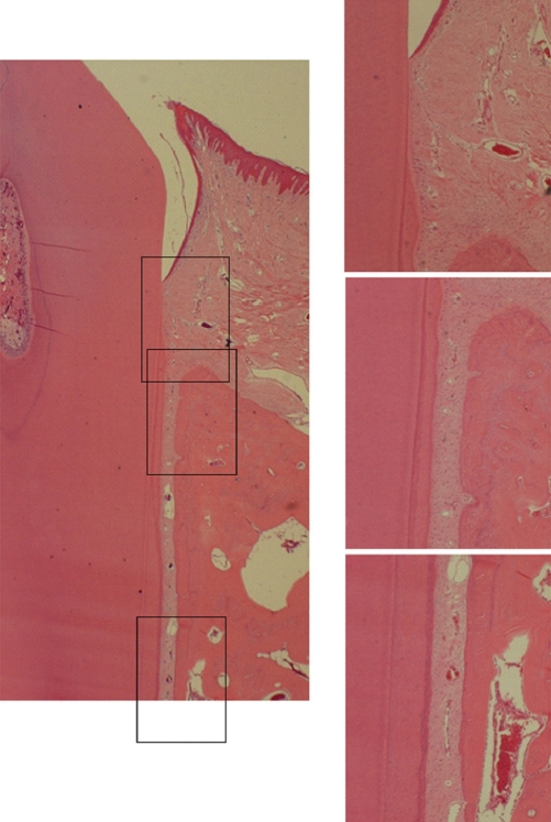
Results: The repaired cementum was composed mostly of acellular cementum and cellular mixed fiber cementum and was thicker in the apical area than in the coronal area. The acellular cementum of the supracrestal area appeared to be amorphous. The newly formed cellular cementum was partially detached from the underlying circumpulpal dentin, which implied a weak attachment between new cementum and dentin, and this split was observed to a lesser extent in the 24 week group than in the 8 week group. The vertical height of the repaired cementum was greater in the 24 week group than in the 8 week group.
Conclusions: Within the limitations of this study, we can conclude that repaired cementum after root planing was mainly acellular cementum and cementum tissue that matured to a shape similar to pristine cementum as the healing progressed from 8 to 24 weeks.
Keywords: Animal models; Dental cementum; Periodontal guided tissue regeneration.
Conflict of interest statement
No potential conflict of interest relevant to this article was reported.
Figures


Similar articles
-
Periodontal repair in surgically created intrabony defects in dogs: influence of the number of bone walls on healing response.J Periodontol. 2004 Feb;75(2):229-35. doi: 10.1902/jop.2004.75.2.229. J Periodontol. 2004. PMID: 15068110
-
GTR treatment of degree III furcation defects following application of enamel matrix proteins. An experimental study in dogs.J Clin Periodontol. 1998 Jun;25(6):524-30. doi: 10.1111/j.1600-051x.1998.tb02482.x. J Clin Periodontol. 1998. PMID: 9667487
-
The effects of a bioabsorbable barrier membrane containing safflower seed extracts on periodontal healing of 1-wall intrabony defects in beagle dogs.J Periodontol. 2005 Jan;76(1):22-33. doi: 10.1902/jop.2005.76.1.22. J Periodontol. 2005. PMID: 15830634
-
Growth/differentiation factor-5 significantly enhances periodontal wound healing/regeneration compared with platelet-derived growth factor-BB in dogs.J Clin Periodontol. 2010 Aug 1;37(8):739-46. doi: 10.1111/j.1600-051X.2010.01576.x. Epub 2010 Jul 1. J Clin Periodontol. 2010. PMID: 20618546
-
Regenerative benefits of using growth factors in treatment of periodontal defects: A systematic review and meta-analysis with Trial Sequential Analysis on preclinical studies.J Tissue Eng Regen Med. 2021 Nov;15(11):964-997. doi: 10.1002/term.3241. Epub 2021 Sep 14. J Tissue Eng Regen Med. 2021. PMID: 34480421
Cited by
-
Visualizing Different Crystalline States during the Infrared Imaging of Calcium Phosphates.Vib Spectrosc. 2020 May;108:103045. doi: 10.1016/j.vibspec.2020.103045. Epub 2020 Feb 24. Vib Spectrosc. 2020. PMID: 35360824 Free PMC article.
-
Effects of vascular formation during alveolar bone process morphogenesis in mice.Histochem Cell Biol. 2017 Oct;148(4):435-443. doi: 10.1007/s00418-017-1584-2. Epub 2017 Jun 13. Histochem Cell Biol. 2017. PMID: 28612087
References
-
- Karring T, Nyman S, Gottlow J, Laurell L. Development of the biological concept of guided tissue regeneration: animal and human studies. Periodontol 2000. 1993;1:26–35. - PubMed
-
- Bartold PM, McCulloch CA, Narayanan AS, Pitaru S. Tissue engineering: a new paradigm for periodontal regeneration based on molecular and cell biology. Periodontol 2000. 2000;24:253–269. - PubMed
-
- Denton GB. The discovery of cementum. J Dent Res. 1939;18:239. (abstract 6). In Robinson HBG. International Association for Dental Research: Proceedings of the Seventeenth General Meeting Hotel Cleveland, Cleveland, Ohio March 18 and 19, 1939. J Dent Res 1939;18:213-303.
-
- Bosshardt DD, Nanci A. Immunodetection of enamel- and cementum-related (bone) proteins at the enamel-free area and cervical portion of the tooth in rat molars. J Bone Miner Res. 1997;12:367–379. - PubMed
-
- Bosshardt DD, Selvig KA. Dental cementum: the dynamic tissue covering of the root. Periodontol 2000. 1997;13:41–75. - PubMed
LinkOut - more resources
Full Text Sources
