

The influence of diabetes mellitus on periodontal tissues: a pilot study
- PMID: 20498760
- PMCID: PMC2872813
- DOI: 10.5051/jpis.2010.40.2.49
The influence of diabetes mellitus on periodontal tissues: a pilot study
Abstract
Purpose: The purpose of this study was to preliminarily evaluate the influence of diabetes mellitus (DM) on periodontal tissue without establishment of periodontitis.
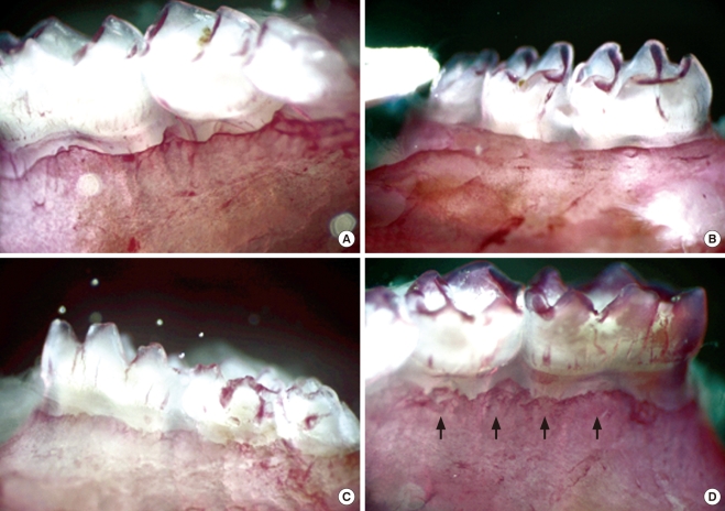
Methods: Seven-week-old db/db mice were used for the diabetic experimental group and systematically healthy mice of the same age were used as controls. After 1 week of acclimatization, the animals were sacrificed for hard and soft tissue evaluation. The pattern of bone destruction was evaluated by stereomicroscope evaluation with alizarin red staining and radiographic evaluation by microscopic computerized tomography images. Histological evaluation was performed with hematoxylin and eosin stain for evaluation of soft tissue changes.
Results: In both stereomicroscope evaluation and radiograph image analysis, aggressive form of bone destruction was observed in diabetic animals when compared to the systematically healthy controls. In histological evaluation, apical migration of junctional epithelium with slight inflammatory cell infiltration was observed with disarrangement of connective tissue fibers.
Conclusions: Within the limits of this study, diabetic animals presented distortion in periodontal attachment and an aggressive bone loss pattern when compared to the healthy controls, suggesting that DM has an independent effect on periodontal tissue destruction irrespective of the presence or absence of periodontal disease.
Keywords: Diabetes mellitus; Inflammation; Periodontal disease.
Conflict of interest statement
No potential conflict of interest relevant to this article was reported.
Figures


Similar articles
-
The influence of diabetes mellitus on periodontal tissues: a histological study.Rom J Morphol Embryol. 2012;53(3):491-5. Rom J Morphol Embryol. 2012. PMID: 22990538
-
Healing of human intrabony defects following regenerative periodontal therapy with a bovine-derived xenograft and guided tissue regeneration.Clin Oral Investig. 2004 Jun;8(2):70-4. doi: 10.1007/s00784-004-0254-7. Epub 2004 Feb 6. Clin Oral Investig. 2004. PMID: 14767696 Clinical Trial.
-
Diabetic Rats Present High Mean Platelet Count in the Presence of Oral Infections.Braz Dent J. 2017 Sep-Oct;28(5):548-551. doi: 10.1590/0103-6440201701386. Braz Dent J. 2017. PMID: 29215677
-
[The relationship of periodontitis and diabetes mellitus].Acta Med Croatica. 2007 Sep;61(4):369-74. Acta Med Croatica. 2007. PMID: 18044471 Review. Croatian.
-
The relationship between diabetes mellitus and destructive periodontal disease: a meta-analysis.Oral Health Prev Dent. 2009;7(2):107-27. Oral Health Prev Dent. 2009. PMID: 19583037 Review.
Cited by
-
Impact of resolvin E1 on murine neutrophil phagocytosis in type 2 diabetes.Infect Immun. 2015 Feb;83(2):792-801. doi: 10.1128/IAI.02444-14. Epub 2014 Dec 8. Infect Immun. 2015. PMID: 25486994 Free PMC article.
-
Chronic Periodontitis in Type 2 Diabetes Mellitus: Oxidative Stress as a Common Factor in Periodontal Tissue Injury.J Clin Diagn Res. 2016 Apr;10(4):BC12-6. doi: 10.7860/JCDR/2016/17350.7542. Epub 2016 Apr 1. J Clin Diagn Res. 2016. PMID: 27190790 Free PMC article.
-
Epigenetic changes caused by diabetes and their potential role in the development of periodontitis.J Diabetes Investig. 2021 Aug;12(8):1326-1335. doi: 10.1111/jdi.13477. Epub 2021 Jan 26. J Diabetes Investig. 2021. PMID: 33300305 Free PMC article.
-
Management of diabolical diabetes mellitus and periodontitis nexus: Are we doing enough?World J Diabetes. 2016 Feb 25;7(4):50-66. doi: 10.4239/wjd.v7.i4.50. World J Diabetes. 2016. PMID: 26962409 Free PMC article. Review.
-
Bone Tissue Engineering (BTE) of the Craniofacial Skeleton, Part II: Translational Potential of 3D-Printed Scaffolds for Defect Repair.J Craniofac Surg. 2024 Jan-Feb 01;35(1):261-267. doi: 10.1097/SCS.0000000000009635. Epub 2023 Aug 25. J Craniofac Surg. 2024. PMID: 37622526 Free PMC article.
References
-
- Mealey BL, Ocampo GL. Diabetes mellitus and periodontal disease. Periodontol 2000. 2007;44:127–153. - PubMed
-
- Shlossman M, Knowler WC, Pettitt DJ, Genco RJ. Type 2 diabetes mellitus and periodontal disease. J Am Dent Assoc. 1990;121:532–536. - PubMed
-
- Mealey BL. Periodontal implications: medically compromised patients. Ann Periodontol. 1996;1:256–321. - PubMed
-
- Papapanou PN. Periodontal diseases: epidemiology. Ann Periodontol. 1996;1:1–36. - PubMed
-
- Emrich LJ, Shlossman M, Genco RJ. Periodontal disease in non-insulin-dependent diabetes mellitus. J Periodontol. 1991;62:123–131. - PubMed
LinkOut - more resources
Full Text Sources
Miscellaneous
