

A hybrid technique for sinus floor elevation in the severely resorbed posterior maxilla
- PMID: 20498764
- PMCID: PMC2872818
- DOI: 10.5051/jpis.2010.40.2.76
A hybrid technique for sinus floor elevation in the severely resorbed posterior maxilla
Abstract
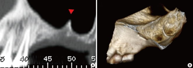
Purpose: This study aimed to evaluate the effectiveness of the modified sinus floor elevation technique described hereafter as a "hybrid technique," in 11 patients with severely resorbed posterior maxillae.
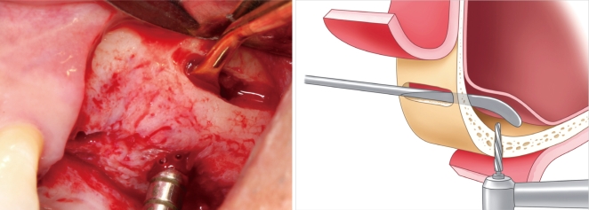
Methods: Eleven patients who received 22 implants in the maxillary premolar and molar areas by the hybrid technique were enrolled in this study. A slot-shaped osteotomy for access was prepared on the lateral wall along the lower border of the sinus floor. The Schneiderian membrane was fully reflected through the lateral slot. Following drilling with the membrane protected by a periosteal elevator, the bone was grafted. All implants were placed simultaneously with sinus augmentation. The cumulative success rate was calculated and clinical parameters were recorded. Radiographic measurements were performed.
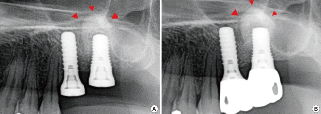
Results: All implants were well maintained at last follow up (cumulative success rate=100%). The mean residual bone height, augmented bone height, crown-to-implant ratio, and marginal bone loss were 4.1+/-1.64 mm, 8.76+/-1.77 mm, 1.21+/-0.33 mm, and 0.34+/-0.72 mm, respectively.
Conclusions: Simultaneous implant placement with sinus augmentation by hybrid technique showed successful clinical results over a 2-year observation period and may be a reliable modality for reconstruction of a severely resorbed posterior maxilla.
Keywords: Bone substitutes; Dental implants; Maxillary sinus.
Conflict of interest statement
No potential conflict of interest relevant to this article was reported.
Figures


References
-
- Jaffin RA, Berman CL. The excessive loss of Branemark fixtures in type IV bone: a 5-year analysis. J Periodontol. 1991;62:2–4. - PubMed
-
- Chanavaz M. Anatomy and histophysiology of the periosteum: quantification of the periosteal blood supply to the adjacent bone with 85Sr and gamma spectrometry. J Oral Implantol. 1995;21:214–219. - PubMed
-
- Sharan A, Madjar D. Maxillary sinus pneumatization following extractions: a radiographic study. Int J Oral Maxillofac Implants. 2008;23:48–56. - PubMed
-
- Pietrokovski J, Massler M. Alveolar ridge resorption following tooth extraction. J Prosthet Dent. 1967;17:21–27. - PubMed
-
- Boyne PJ, James RA. Grafting of the maxillary sinus floor with autogenous marrow and bone. J Oral Surg. 1980;38:613–616. - PubMed
LinkOut - more resources
Full Text Sources
