

Characterization of tumor progression in engineered tissue using infrared spectroscopic imaging
- PMID: 20498913
- PMCID: PMC3030988
- DOI: 10.1039/c0an00112k
Characterization of tumor progression in engineered tissue using infrared spectroscopic imaging
Abstract
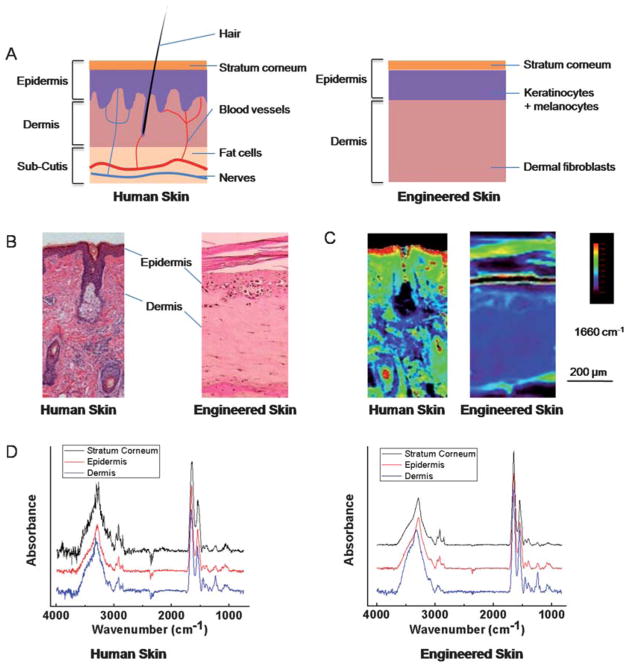
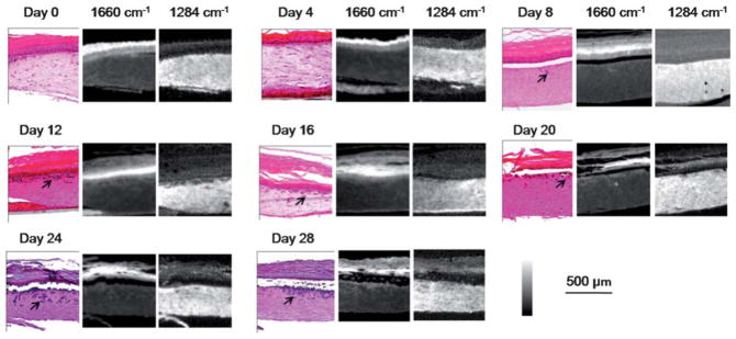
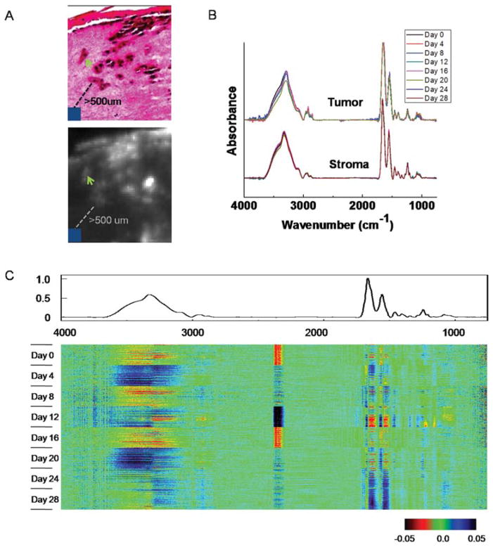
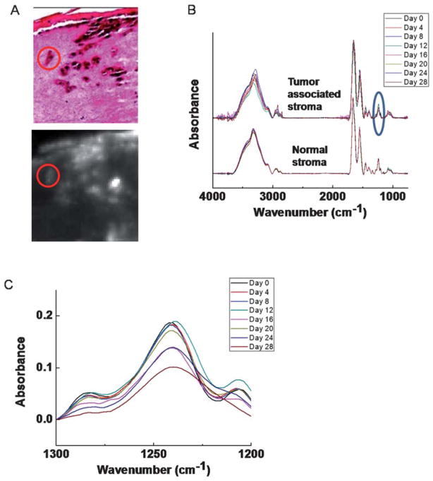
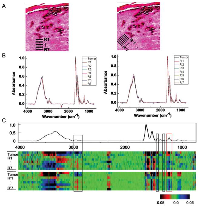
Engineered tissues can provide models for imaging and disease progression and the use of such models is becoming increasingly prevalent. While structural characterization of these systems is documented, a combination of biochemical and structural knowledge is often helpful. Here, we apply Fourier transform infrared (FT-IR) spectroscopic imaging to examine an engineered tissue model of melanoma. We first characterize the biochemical properties and spectral changes in different layers of growing skin. Second, we introduce malignant melanocytes to simulate tumor formation and growth. Both cellular changes associated with tumor formation and growth can be observed. In particular, chemical changes associated with tumor-stromal interactions are observed during the course of tumor growth and appear to influence a 50-100 microm region. The development of this analytical approach combining engineered tissue with spectroscopy, imaging and computation will allow for quality control and standardization in tissue engineering and novel scientific insight in cancer progression.
Figures

References
-
- Melanoma Study Group of the Mayo Clinic Cancer Center. Mayo Clin Proc. 2007;82:364–380. - PubMed
-
- Thompson JF, Scolyer RA, Kefford RF. Lancet. 2005;365:687–701. - PubMed
-
- James WD, Berger TG, Elston D. Andrews’ Diseases of the Skin. 9. Philadelphia: 2000. pp. 881–889.
-
- Elder DE. Clin Cancer Res. 2006;12:2308s–2311s. - PubMed
-
- Hsu MY, Meier F, Herlyn M. Differentiation. 2002;70:522–536. - PubMed
Publication types
MeSH terms
Grants and funding
LinkOut - more resources
Full Text Sources
Medical