

Specimen-specific method for quantifying glenohumeral joint kinematics
- PMID: 20499181
- PMCID: PMC2940013
- DOI: 10.1007/s10439-010-0074-7
Specimen-specific method for quantifying glenohumeral joint kinematics
Abstract
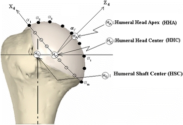
The existing glenohumeral joint kinematic protocols are highly effective for studying in vivo shoulder kinematics but are not anatomically specific enough to address the asymmetric changes in glenohumeral joint kinematics and do not provide clear anatomic definitions for landmarks and directions. Therefore, the objective of this study was to develop an anatomically relevant and specimen-specific three-dimensional glenohumeral joint kinematic method as a new standard definition protocol for the glenohumeral coordinate systems (CSs). The in situ kinematic data of the intra-capsular glenoid-based CS of the glenohumeral joint were mathematically determined from the kinematic data of the extra-capsular CSs measured with an intact capsule. To minimize irreproducibility arising from discrepancy in initial specimen condition and error in determining CSs, several techniques were employed to determine anatomical landmarks and directions. To examine and demonstrate the details of this method, six fresh frozen cadaveric shoulders were used with a custom shoulder testing system. The accuracy and repeatability in the humeral head center (HHC) measurement were 0.44 and 0.41 mm, respectively. The inter-observer reliability for the location of the glenoid CS origin and HHC were 0.37 and 0.30 mm, respectively. The smaller anteroposterior (AP) depth of the glenoid with respect to the superoinferior (SI) depth (27.3 ± 16.5%) was significantly correlated to the larger AP/SI translation ratio of the humeral head apex (191.4 ± 43.8%, R = 0.90, p = 0.02). This study provides a glenohumeral kinematic protocol that enables the assessment of asymmetric glenohumeral kinematics determined by a precise and reproducible method using anatomic landmarks.
Figures


References
-
- Beaulieu CF, Hodge DK, Bergman AG, Butts K, Daniel BL, Napper CL, Darrow RD, Dumoulin CL, Herfkens RJ. Glenohumeral relationships during physiologic shoulder motion and stress testing: initial experience with open MR imaging and active imaging-plane registration. Radiology. 1999;212:699–705. - PubMed
