. 2010 Oct;17(10):2619-27.
doi: 10.1245/s10434-010-1121-1. Epub 2010 May 25.

Higher expression of the heterogeneous nuclear ribonucleoprotein k in melanoma

Affiliations

Higher expression of the heterogeneous nuclear ribonucleoprotein k in melanoma

Fushi Wen et al. Ann Surg Oncol. 2010 Oct.

Abstract

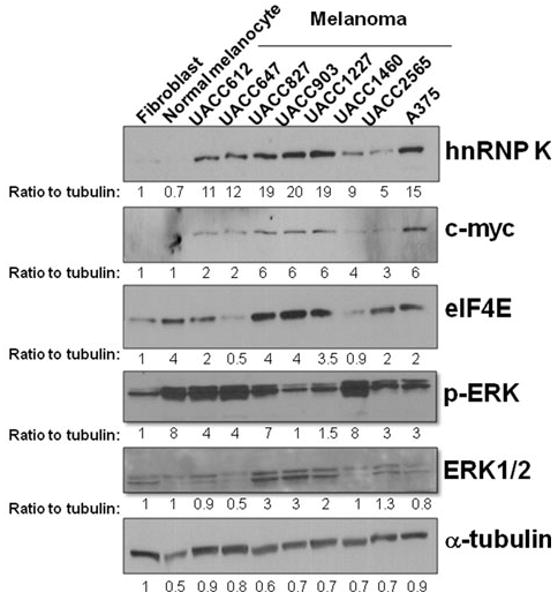
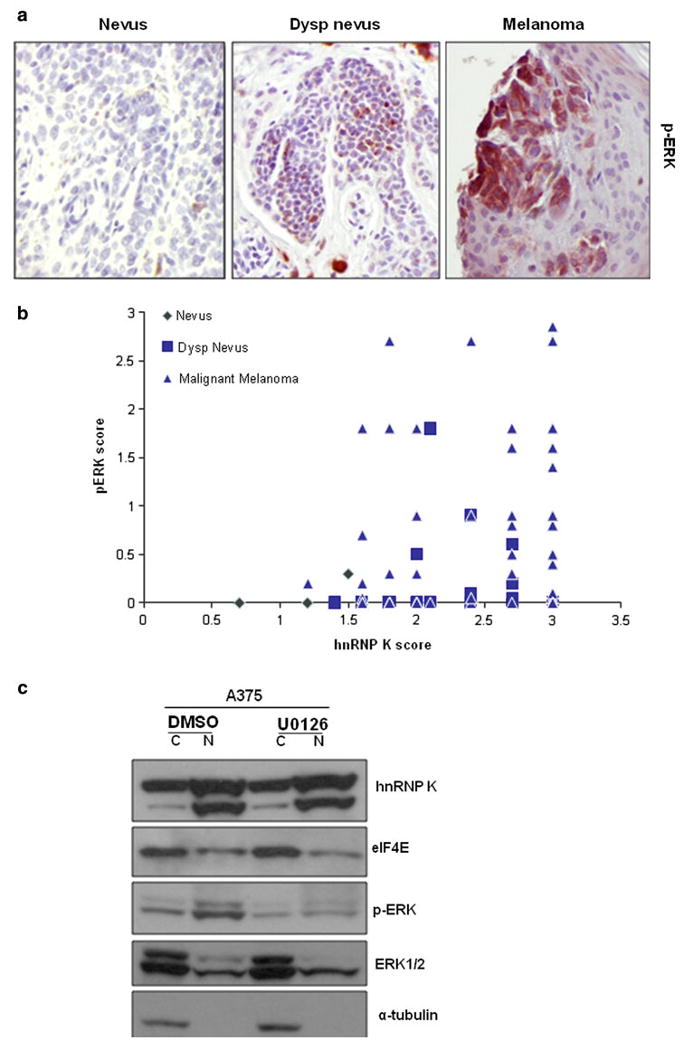
Background: The heterogeneous nuclear ribonucleoprotein (hnRNP) K is an essential RNA and DNA binding protein involved in gene expression and signal transduction. The role of hnRNP K in cancer is relatively understudied. However, several cellular functions strongly indicate that hnRNP K is involved in tumorigenesis. Oncogenes c-Src, c-myc, and eIF4E are regulated by hnRNP K. We have shown an increased cytoplasmic hnRNP K in pancreatic cancer. In the present study, we investigated the altered expression of hnRNP K protein and its correlation with p-ERK in melanoma using human melanoma cell lines and tissue microarray.

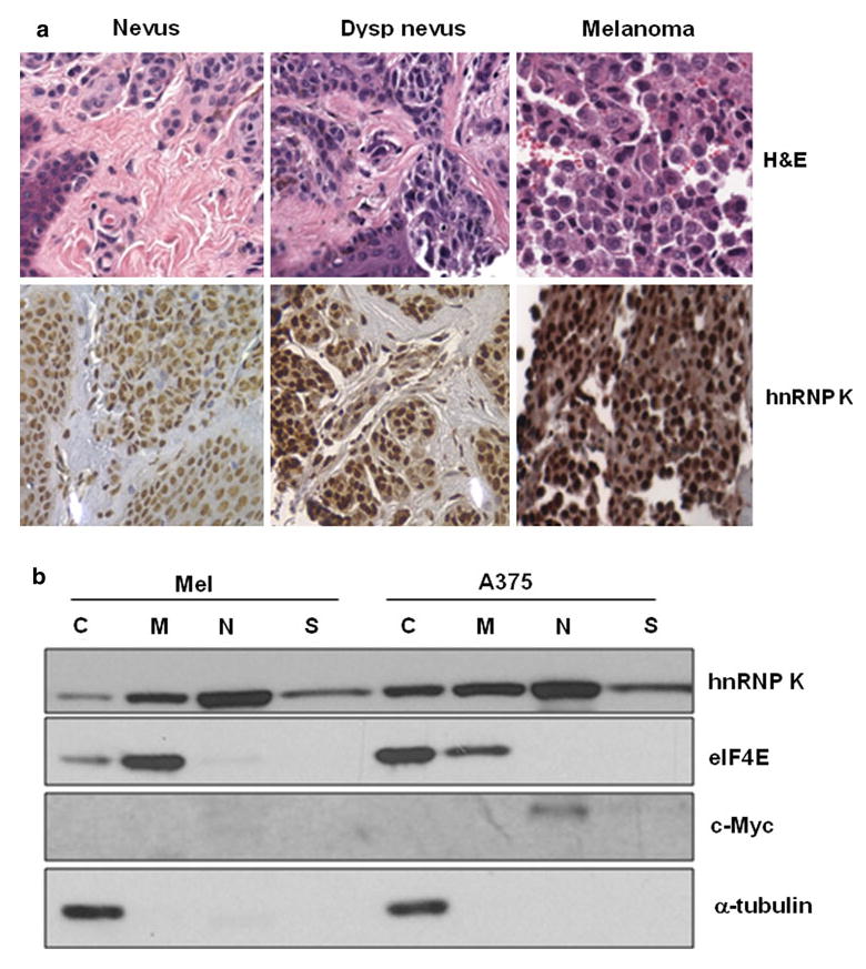
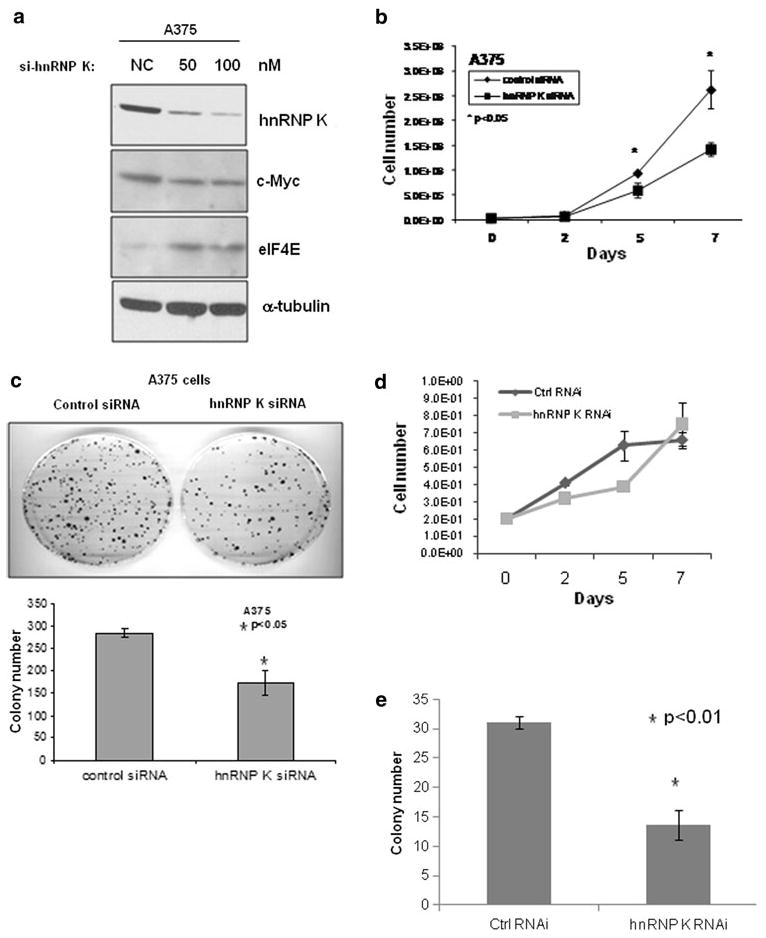
Materials and methods: The protein levels of hnRNP K and p-ERK in 8 human melanoma cell lines and a melanoma progression tissue microarray containing 80 melanoma, 23 dysplastic nevi, and 14 benign nevi specimens were analyzed using Western blot and immunohistochemistry analysis. hnRNP K was knocked down by siRNA, and its effect on melanoma cells was assessed.

Results: We showed a higher hnRNP K protein level in both melanoma cell lines and melanoma tissue specimens, which correlated with a higher c-myc expression. An increase in the cytoplasmic hnRNP K and eIF4E protein levels in melanoma cells is also seen. p-ERK level was also higher in dysplastic nevi and melanoma tissues, but did not correlate with hnRNP K protein level. We then demonstrated that knocking down of hnRNP K by siRNA inhibited melanoma cell growth and colony formation, as well as c-myc expression.

Conclusions: hnRNP K expression correlated with melanoma and may play a role in melanoma tumorigenesis.


Figures

FIG. 1
FIG. 1
Higher hnRNP K protein level in human melanoma cell lines. Primary fibroblast, primary normal melanocytes, and 8 melanoma cell lines were grown at the log phase and harvested. Western blot analysis was performed using hnRNP K, c-myc, eIF4E, p-ERK, ERK1/2, and α-tubulin antibodies. The intensities of the bands were quantified using ImageJ software and normalized to α-tubulin. The ratio to tubulin was indicated below each band. ERK1/2 was measured as 1 band because of the close distance between these 2 bands
FIG. 2
FIG. 2
a hnRNP K protein is increased in human dysplastic nevus and melanoma tissues. H&E staining and immunohistochemistry was performed on a melanoma progression tissue microarray using hnRNP K antibody. Representative H&E and immunohistochemistry analysis of the hnRNP K protein in benign nevus, dysplastic nevus, and melanoma tissues were shown. Magnification: 400 ×. b Increased cytoplasmic hnRNP K protein in melanoma cells. Mel-STV (Mel) and A375 cells were fractionated, and Western blot analysis was performed using hnRNP K, eIF4E, c-myc and α-tubulin antibodies. C cytoplasm, M membrane, N nucleus, S cytoskeleton
FIG. 3
FIG. 3
a p-ERK level is higher in human dysplastic nevus and melanoma tissues. Immunohistochemistry was performed on the same melanoma progression tissue microarray using p-ERK antibody. Representative immunohistochemistry analysis of p-ERK in benign nevus, dysplastic nevus, and melanoma tissues were shown. Magnification: 200 ×. b p-ERK level does not correlate with hnRNP K protein level. The IHC scores of the hnRNP K protein level and p-ERK level of the same tissue specimens were plotted against each other. c MEK inhibitor did not decrease the accumulation of hnRNP K and eIF4E in the cytoplasm. A375 cells were treated with MEK inhibitor U0126 (10 μM) or vehicle control DMSO for 1 h and fractionated. Western blot analysis was performed using hnRNP K, eIF4E, p-ERK, ERK1/2, and α-tubulin antibodies. C cytoplasm, N nucleus
FIG. 4
FIG. 4
siRNA knock down of hnRNP K inhibited melanoma cell growth and colony formation. a A375 melanoma cells were transfected with negative control (NC) or 50 nM or 100 nM of hnRNP K siRNAs. Cells were harvested and lysed 48 h after transfection, followed by Western blot analysis using hnRNP K, c-myc, eIF4E, and α-tubulin antibodies. b A375 cells were transfected with 50 nM of negative control or hnRNP K siRNA. Then, 24 h after transfection, 2 × 10⁴ cells were seeded into 100-mm plates in triplicate, and cell growth was monitored by counting total cell numbers every 2 or 3 days; or c 1000 cells were seeded into 100-mm plates in triplicate and incubated for 2 weeks to allow colony formation. Then media were removed, and colonies were stained with methylene blue solution. The plates were rinsed with water, and the colony number was counted. d, e Same experiments as described in b and c were repeated in primary human fibroblasts

References

    1. Bomsztyk K, Denisenko O, Ostrowski J. hnRNP K: one protein multiple processes. Bioessays. 2004;26:629–38. - PubMed
    1. Klimek-Tomczak K, Wyrwicz LS, Jain S, Bomsztyk K, Ostrowski J. Characterization of hnRNP K protein-RNA interactions. J Mol Biol. 2004;342:1131–41. - PubMed
    1. Mikula M, Dzwonek A, Karczmarski J, Rubel T, Dadlez M, Wyrwicz LS, et al. Landscape of the hnRNP K protein-protein interactome. Proteomics. 2006;6:2395–406. - PubMed
    1. Lynch M, Chen L, Ravitz MJ, Mehtani S, Korenblat K, Pazin MJ, et al. hnRNP K binds a core polypyrimidine element in the eukaryotic translation initiation factor 4E (eIF4E) promoter, and its regulation of eIF4E contributes to neoplastic transformation. Mol Cell Biol. 2005;25:6436–53. - PMC - PubMed
    1. Baber JL, Libutti D, Levens D, Tjandra N. High precision solution structure of the C-terminal KH domain of heterogeneous nuclear ribonucleoprotein K, a c-myc transcription factor. J Mol Biol. 1999;289:949–62. - PubMed

Publication types

MeSH terms

Substances