. 2010 Oct;25(10):2175-83.
doi: 10.1002/jbmr.115.

Osteosclerosis owing to Notch gain of function is solely Rbpj-dependent

Affiliations

Osteosclerosis owing to Notch gain of function is solely Rbpj-dependent

Jianning Tao et al. J Bone Miner Res. 2010 Oct.

Abstract

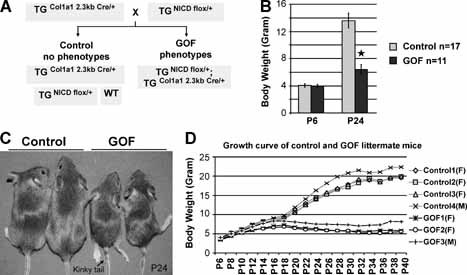
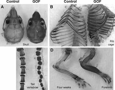
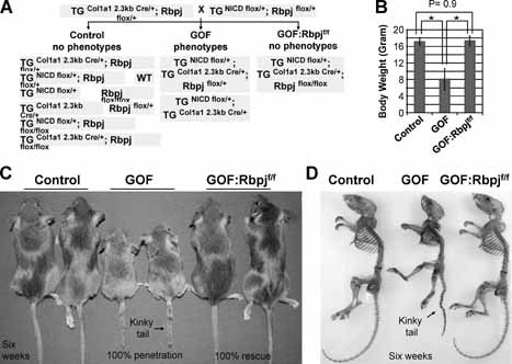
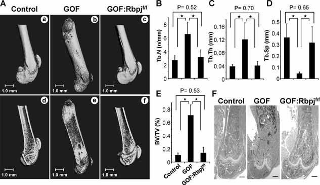
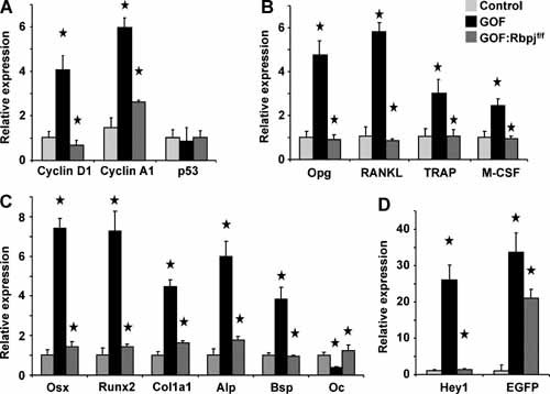
Osteosclerosis is a pathologic bone disease characterized by an increase in bone formation over bone resorption. Genetic factors that contribute to the pathogenesis of this disease are poorly understood. Dysregulation or mutation in many components of the Notch signaling pathway results in a wide range of human developmental disorders and cancers, including bone diseases. Our previous study found that activation of the Notch signaling in osteoblasts promotes cell proliferation and inhibits differentiation, leading to an osteosclerotic phenotype in transgenic mice. In this study we report a longer-lived mouse model that also develops osteosclerosis and a genetic manipulation that completely rescues the phenotype. Conditionally cre-activated expression of Notch1 intracellular domain (NICD) in vivo exclusively in committed osteoblasts caused massive osteosclerosis with growth retardation and abnormal vertebrae. Importantly, selective deletion of a Notch nuclear effector--Rbpj--in osteoblasts completely suppressed the osteosclerotic and growth-retardation phenotypes. Furthermore, cellular and molecular analyses of bones from the rescued mice confirmed that NICD-dependent molecular alterations in osteoblasts were completely reversed by removal of the Rbpj pathway. Together, our observations show that the osteosclerosis owing to activation of Notch signaling in osteoblasts is canonical in nature because it depends solely on Rbpj signaling. As such, it identifies Rbpj as a specific target for manipulating Notch signaling in a cell-autonomous fashion in osteoblasts in bone diseases where Notch may be dysregulated.


Figures

Figure 1
Figure 1
Cre recombinase‐activation of Notch gain of function in bitransgenic mice causes growth retardation and kinky tail phenotypes. (A) Breeding scheme of NICD bitransgenic GOF mice. The transgenic mice (TGNICDflox/+) were generated by Doug Melton using a knock‐in allele of NICD cDNA with a flox/STOP cassette inserted into the Rosa26 locus. Col1a1 2.3‐kb Cre that is specifically expressed in osteoblasts will activate transcription of single‐copy NICD in GOF mice to bypass lethality of traditional transgenic over expression of NICD in osteoblasts. (B) Body weights of GOF versus control mice on postnatal days 6 (P6) and 24 (P24) show similar weights early but significant differences by weaning (control n = 17, GOF n = 11; *p < .001). (C) Mice on postnatal day 24 (P24) with control pair and GOF pair. The latter displays smaller body size and kinky tails (black arrow). (D) Representative growth curves of four control mice and three GOF littermate mice from P6 to P40. Body weights were measured every other day. Both GOF females (F) and males (M) grow more slowly than control littermates commencing at or near 2 weeks of age.
Figure 2
Figure 2
Notch gain of function causes generalized osteosclerosis in bitransgenic mice. Skeletal preparation of 4‐week‐old GOF mice showed thickened bones of a severe osteosclerotic phenotype in skull (A), ribs (B), tail vertebrae (C), and forelimb (D). Skeletal preparations were stained with alcian blue and alizarin red.
Figure 3
Figure 3
Genetic addition of the Rbpjflox/flox allele rescues growth retardation and kinky‐tail phenotypes in bitransgenic mice. (A) Breeding scheme of NICD bitransgenic GOF mice with Rbpjflox/flox mice. These GOF:Rbpjf/f mice have an activating NICD allele and deletion of Rbpj in committed osteoblasts. (B) Examination shows that GOF mice at 6 weeks of age have significantly smaller body weight than control and GOF:Rbpjf/f mice (control n = 12, GOF n = 8, GOF:Rbpjf/f n = 3). The GOF:Rbpjf/f mice have similar weights as controls. *p < .001 between control and GOF mice as well as between GOF and GOF:Rbpjf/f mice. (C) Mice at 6 weeks of age with control pair, GOF pair, and GOF:Rbpjf/f pair. Only the GOF pair displays smaller body size and kinky tail (black arrow). (D) Skeletal preparations of 6‐week‐old GOF:Rbpjf/f mice show the normal skeletal shape and size of the GOF:Rbpjf/f skeleton with a straight tail. Control, GOF, GOF:Rbpjf/f mice are littermates. Skeletal preparations were stained with alcian blue and alizarin red.
Figure 4
Figure 4
The osteosclerotic phenotype in the Notch GOF mice is reversible. (A) µCT reconstruction of distal (a, c) or whole femurs (b) from 2‐month‐old mice shows a shortening in GOF mice compared with control or GOF:Rbpjf/f mice. (d–f) A sagittal section of femur from (a–c). Trabecular bone in GOF mice is increased (e), and this change is decreased to normal in GOF:Rbpjf/f mice (f) compared with control (d). Scale bar = 1.0 mm. (B–E) Quantitative bone histomorphometric analyses of the femurs in 2‐month‐old control, GOF, and GOF:Rbpjf/f mice (n = 5). The increased trabecular number (B), thickness (C), and bone volume/tissue volume ratio (BV/TV, E) and decreased trabecular spacing (D) in the GOF mice are reversed in the GOF:Rbpjf/f mice (*p < .05 groups). (F) Goldner's staining of the distal femurs from 2‐month‐old mice shows that the increased trabecular bone in the GOF mice (middle panel) is decreased on the Rbpjf/f background (right panel) to a level similar to control (left panel). Scale bars = 500 µm.
Figure 5
Figure 5
The molecular signature of osteosclerosis is reversed in the GOF:Rbpjf/f osteoblasts. Calvarial total RNA was obtained from control, GOF, and GOF:Rbpjf/f mice at 3 weeks of age (n = 4). The transcriptional profile for cell cycle, osteoblast, and osteoclastic markers was rescued: (A) qRT‐PCR of cell cycle markers cyclin D1, cyclin A1, and p53; (B) qRT‐PCR of macrophage and osteoclast differentiation markers osteoprotegerin, RANKL, RANK ligand, TRACP, and M‐CSF; (C) qRT‐PCR of early and late osteoblastic differentiation markers osterix, Runx2, Col1a1, ALP, BSP, and osteocalcin; (D) qRT‐PCR of the Notch target gene Hey1 and transgene marker EGFP. *p < .05 between control and GOF as well as between GOF and GOF:Rbpjf/f.

References

    1. Whyte MP. Sclerosing bone disorders In: Rosen CJ, ed. Primer on the Metabolic Bone Diseases and Disorders of Mineral Metabolism, 7th ed Washington, DC: American Society for Bone and Mineral Research, 2008: 412–423.
    1. De Vernejoul MC. Sclerosing bone disorders. Best Pract Res Clin Rheumatol. 2008; 22: 71–83. - PubMed
    1. Kurland ES, Schulman RC, Zerwekh JE, Reinus WR, Dempster DW, Whyte MP. Recovery from skeletal fluorosis (an enigmatic American case). J Bone Miner Res. 2007; 22: 163–170. - PubMed
    1. Whyte MP, Totty WG, Lim VT, Whitford GM. Skeletal fluorosis from instant tea. J Bone Miner Res. 2008; 23: 759–769. - PubMed
    1. Chavassieux P, Seeman E, Delmas PD. Insights into material and structural basis of bone fragility from diseases associated with fractures: how determinants of the biomechanical properties of bone are compromised by disease. Endocr Rev. 2007; 28: 151–164. - PubMed

Publication types

MeSH terms

Substances