

Peak bone mass from longitudinal data: implications for the prevalence, pathophysiology, and diagnosis of osteoporosis
- PMID: 20499378
- PMCID: PMC5101070
- DOI: 10.1002/jbmr.95
Peak bone mass from longitudinal data: implications for the prevalence, pathophysiology, and diagnosis of osteoporosis
Abstract
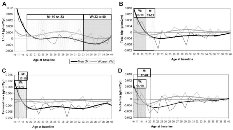
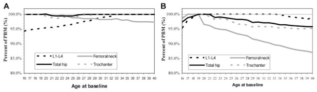
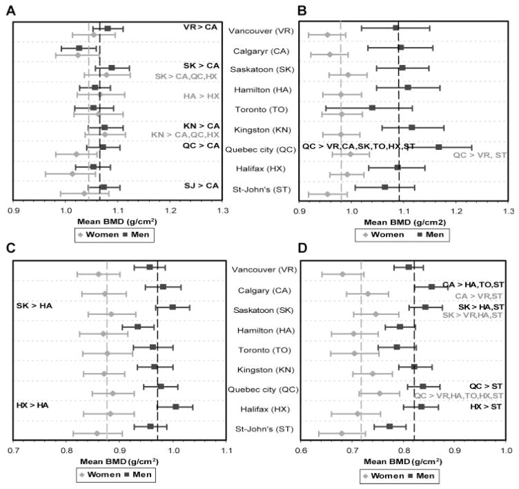
We estimated peak bone mass (PBM) in 615 women and 527 men aged 16 to 40 years using longitudinal data from the Canadian Multicentre Osteoporosis Study (CaMos). Individual rates of change were averaged to find the mean rate of change for each baseline age. The age range for PBM was defined as the period during which bone mineral density (BMD) was stable. PBM was estimated via hierarchical models, weighted according to 2006 Canadian Census data. Lumbar spine PBM (1.046 ± 0.123 g/cm(2)) occurred at ages 33 to 40 years in women and at 19 to 33 years in men (1.066 ± 0.129 g/cm(2)). Total hip PBM (0.981 ± 0.122 g/cm(2)) occurred at ages 16 to 19 years in women and 19 to 21 years in men (1.093 ± 0.169 g/cm(2)). Analysis of Canadian geographic variation revealed that the levels of PBM and of mean BMD in those over age 65 sometimes were discordant, suggesting that PBM and subsequent rates of bone loss may be subject to different genetic and/or environmental influences. Based on our longitudinally estimated PBM values, the estimated Canadian prevalences of osteoporosis (T-score < -2.5) were 12.0% (L(1)-L(4)) and 9.1% (total hip) in women aged 50 years and older and 2.9% (L(1)-L(4)) and 0.9% (total hip) in men aged 50 years and older. These were higher than prevalences using cross-sectional PBM data. In summary, we found that the age at which PBM is achieved varies by sex and skeletal site, and different reference values for PBM lead to different estimates of the prevalence of osteoporosis. Furthermore, lack of concordance of PBM and BMD over age 65 suggests different determinants of PBM and subsequent bone loss.
© 2010 American Society for Bone and Mineral Research.
Figures
References
-
- Report of a WHO Study Group. World Health Organ Techical Report Series 843. Geneva: WHO; 1994. Assessment of Fracture Risk and Its Application to Screening for Postmenopausal Osteoporosis; pp. 1–129. - PubMed
-
- Pedrazzoni M, Girasole G, Bertoldo F, et al. Definition of a population-specific DXA reference standard in Italian women: the Densitometric Italian Normative Study (DINS) Osteoporos Int. 2003;14:978–982. - PubMed
-
- Hoiberg M, Nielsen TL, Wraae K, et al. Population-based reference values for bone mineral density in young men. Osteoporos Int. 2007;18:1507–1514. - PubMed
-
- Bachrach LK, Hastie T, Wang MC, Narasimhan B, Marcus R. Bone mineral acquisition in healthy Asian, Hispanic, black, and Caucasian youth: a longitudinal study. J Clin Endocrinol Metab. 1999;84:4702–4712. - PubMed
-
- Looker AC, Wahner HW, Dunn WL, et al. Updated data on proximal femur bone mineral levels of US adults. Osteoporos Int. 1998;8:468–489. - PubMed
Publication types
MeSH terms
Grants and funding
LinkOut - more resources
Full Text Sources
Medical
