. 2010 Jul;19(7):1420-31.
doi: 10.1002/pro.423.

Automated electron-density sampling reveals widespread conformational polymorphism in proteins

Affiliations

Automated electron-density sampling reveals widespread conformational polymorphism in proteins

P Therese Lang et al. Protein Sci. 2010 Jul.

Abstract

Although proteins populate large structural ensembles, X-ray diffraction data are traditionally interpreted using a single model. To search for evidence of alternate conformers, we developed a program, Ringer, which systematically samples electron density around the dihedral angles of protein side chains. In a diverse set of 402 structures, Ringer identified weak, nonrandom electron-density features that suggest of the presence of hidden, lowly populated conformations for >18% of uniquely modeled residues. Although these peaks occur at electron-density levels traditionally regarded as noise, statistically significant (P < 10(-5)) enrichment of peaks at successive rotameric chi angles validates the assignment of these features as unmodeled conformations. Weak electron density corresponding to alternate rotamers also was detected in an accurate electron density map free of model bias. Ringer analysis of the high-resolution structures of free and peptide-bound calmodulin identified shifts in ensembles and connected the alternate conformations to ligand recognition. These results show that the signal in high-resolution electron density maps extends below the traditional 1 sigma cutoff, and crystalline proteins are more polymorphic than current crystallographic models. Ringer provides an objective, systematic method to identify previously undiscovered alternate conformations that can mediate protein folding and function.


Figures
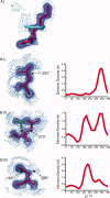
Figure 1
Figure 1
Ringer systematically samples electron density in real space. (A) Ringer computes electron density (1.0 σ, solid; 0.3 σ, mesh) around idealized dihedral angles (cyan ring) extending from the modeled side chain (red). Shown here is Lys86 from a putative tyrosine phosphatase from Rhodopseudomonas palustris (PDB ID 2HHG). (B) Ringer identifies several types of conformational polymorphism in the 1.0 Å-resolution electron density of oxy-myoglobin (PDB ID 1A6M26). Left: the model is superimposed on the electron density map displayed at the standard cutoff (1.0 σ, solid) and the mean electron density (0.0 σ, mesh). Right: plots of electron density (σ) as a function of χ1 angle. (I) Asp27: a single peak in the electron density distribution around a uniquely modeled residue. (II) Glu4: multiple peaks from multiple conformations (red, major; green, minor) in the model. (III) Asn132: multiple peaks in the electron density interpreted with a unique conformation in the model.
Figure 2
Figure 2
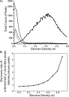
Analysis of a test set of 402 high-resolution structures suggests a lower threshold for defining side-chain electron density. (A) Distribution of Ringer peak heights at the C–C bond length for primary (solid), secondary (short dash), and tertiary peaks (dash) for residues that are unbranched at χ1. In comparison to the alanine primary electron density peak heights (dot), also sampled at C–C bond length, the secondary and tertiary peaks of longer side chains are enriched above 0.3 σ. (B) Lower electron density threshold that enriches for heavy-atom over hydrogen contributions at the C–C bond length. Discovery rate, calculated as the ratio of the number χ1 secondary electron density peaks (normalized by the total number of χ1 side chains) to alanine primary electron density peaks (normalized by the total number of alanines), is plotted versus the lower electron density cutoff (σ).
Figure 3
Figure 3
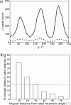
Weak electron-density features are enriched in rotameric positions. (A) Distribution of secondary peaks (solid line) ≥0.3 σ versus χ1 angle shows a trimodal distribution strongly enriched for preferred rotameric positions. The distribution of tertiary peaks (dotted line) ≥0.3σ shows a similar tri-modal distribution. In contrast, the distribution of quaternary peaks, which cannot physically correspond to chemical features of the side chains, is random (data not shown). (B) Percent of the total secondary peaks within the indicated angular difference from ideal rotameric values. Peaks are enriched over a random distribution (dashed line) up to 30° from the rotameric values.
Figure 4
Figure 4
Ringer detects peaks at correlated χ1 and χ2 angles. (A) Peaks in electron density (1.0 σ, solid; 0.3 σ, mesh) are identified by sampling χ1 (cyan ring) and then sampling χ2 (purple ring) at idealized bond lengths from the χ1 secondary peak (orange sphere). (B) A histogram of secondary χ1 peaks and primary χ2 peaks built from the unmodeled χ1 peaks. Cross-peaks are significantly enriched in rotameric positions (P-value < 10−5).
Figure 5
Figure 5
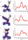
Ringer detects evidence for minor unmodeled conformations in an electron density map calculated with accurate experimental phases. Ringer plots of electron density (σ) versus χ1 angle for three representative residues in the RH4B coiled coil (PDB ID 2O6N31). The orange line indicates 0.3 σ. The experimental, MAD-phased electron density at 1 σ (solid) and 0.3 σ (mesh) is shown with the modeled conformations (red) for residues (A) Gln5, (B) Lys7, and (C) Glu20. Structures of the minor rotamers (orange) have been added to guide the eye and are not the result of refinement.
Figure 6
Figure 6
Resolution dependence of Ringer peak detection at the 0.3 σ threshold. (A) Percent of unique residues unbranched at χ1 with secondary peaks ≥0.3 σ over a range of resolutions (white). The percent of residues with modeled alternate conformations is displayed in black. (B) The percentage of secondary Ringer peaks within 30° of rotameric angles over a range of resolutions.
Figure 7
Figure 7
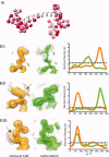
Ringer finds evidence for functionally relevant, unmodeled minor conformers in CaM. (A) Distribution in unbound CaM of unmodeled (red spheres) and modeled alternate conformations (pink spheres). (B) Electron density features of unbound CaM (orange; 1.0 Å resolution) and CaM complexed with the smMLCK peptide (green; 1.08 Å resolution). Modeled conformations are shown superimposed on the electron density at 1 σ (solid) and 0.3 σ (mesh). Arrows indicate areas of interest in electron density. Ringer plots of electron density (σ) versus χ1 angle for three representative residues. Gray dashed line indicates 0.3 σ. (I) Arg126: single peaks ≥0.3 σ in the electron density distribution around each uniquely modeled residue. (II) Ser38: the unbound structure has two electron-density peaks ≥0.3 σ, and the minor peak is strongly enriched in the peptide complex. Selection of the secondary peak supports a population shift upon binding. (III) Asp22: the unbound structure shows two peaks ≥0.3 σ. The secondary peak is absent in the smMLCK peptide complex, suggesting rigidification of this residue accompanies binding.

Comment in

  • Peripatetic proteins.
    Matthews BW. Matthews BW. Protein Sci. 2010 Jul;19(7):1279-80. doi: 10.1002/pro.422. Protein Sci. 2010. PMID: 20499369 Free PMC article. No abstract available.

References

    1. Vendruscolo M. Determination of conformationally heterogeneous states of proteins. Curr Opin Struct Biol. 2007;17:15–20. - PubMed
    1. Frederick KK, Marlow MS, Valentine KG, Wand AJ. Conformational entropy in molecular recognition by proteins. Nature. 2007;448:325–U323. - PMC - PubMed
    1. Boehr DD, McElheny D, Dyson HJ, Wright PE. The dynamic energy landscape of dihydrofolate reductase catalysis. Science. 2006;313:1638–1642. - PubMed
    1. Fraser JS, Clarkson MW, Degnan SC, Erion R, Kern D, Alber T. Hidden alternative structures of proline isomerase essential for catalysis. Nature. 2009;462:669–673. - PMC - PubMed
    1. Li PL, Martins IRS, Amarasinghe GK, Rosen MK. Internal dynamics control activation and activity of the autoinhibited Vav DH domain. Nat Struct Mol Biol. 2008;15:613–618. - PMC - PubMed

Publication types