

Cesium implant for tongue carcinoma with a thickness of 1.5 cm or more: cases successfully treated with a Modified Manchester System
- PMID: 20499422
- PMCID: PMC2880269
- DOI: 10.3349/ymj.2010.51.4.557
Cesium implant for tongue carcinoma with a thickness of 1.5 cm or more: cases successfully treated with a Modified Manchester System
Abstract
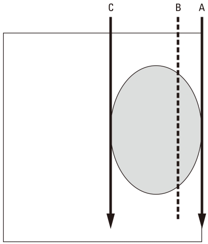
Purpose: Deciding on treatment carcinoma of the tongue when the tumor has a thickness of 1.5 cm or more is difficult. Surgery often requires wide resection and re-construction, leading to considerable functional impairment. A cesium implant is an attractive option, but according to the Manchester System, a two plane implant is needed.
Materials and methods: According to the textbook, a tumor is sandwiched between the needles, which are implanted at the edge of the tumor. This may cause an unnecessarily high dose to the outer surface of the tongue, which sometimes leads to a persistent ulcer. To avoid this complication, we invented a modified implantation method, and applied the method to five consecutive patients.
Results: With a minimum follow-up of 2 years, all primary tumors in 5 consecutive patients have been controlled. No complications occurred in soft tissue of the tongue or in the mandible.
Conclusion: Our modified Manchester System was feasible and effective for tumors that has a thickness of 1.5 cm or more.
Conflict of interest statement
The authors have no financial conflicts of interest.
Figures


References
-
- Duthie MB, Gupta NK, Pointon CS. Head and neck. In: Pointon RCS, editor. The radiotherapy of malignant disease. London: Springer-Verlag; 1991. p. 156.
-
- Hosokawa Y, Shirato H, Nishioka T, Tsuchiya K, Chang TC, Kagei K, et al. Effect of treatment time on outcome of radiotherapy for oral tongue carcinoma. Int J Radiat Oncol Biol Phys. 2003;57:71–78. - PubMed
-
- Mendenhall WM, Riggs CE, Cassisi NJ. Treatment of head and neck cancers. In: Devita VT, Hellman S, Rosenberg S, editors. Cancer principle and practice of oncology. Philadelphia: Lippincott Williams and Wilkins; 2005. p. 682.
-
- Obinata K, Ohmori K, Tuchiya K, Nishioka T, Shirato H, Nakamura M. Clinical study of a spacer to help prevent osteoradionecrosis resulting from brachytherapy for tongue cancer. Oral Surg Oral Med Oral Pathol Oral Radiol Endod. 2003;95:246–250. - PubMed
-
- O'Brien CJ, Lauer CS, Fredricks S, Clifford AR, McNeil EB, Bagia JS, et al. Tumor thickness influences prognosis of T1 and T2 oral cavity cancer--but what thickness? Head Neck. 2003;25:937–945. - PubMed
Publication types
MeSH terms
Substances
LinkOut - more resources
Full Text Sources
