

Finite element analysis of eustachian tube function in cleft palate infants based on histological reconstructions
- PMID: 20500073
- PMCID: PMC3044605
- DOI: 10.1597/09-131
Finite element analysis of eustachian tube function in cleft palate infants based on histological reconstructions
Abstract
Introduction: The prevalence of otitis media with effusion approaches 100% in infants with cleft palate (CP), and disease pathogenesis is believed to be caused by eustachian tube (ET) dysfunction.
Objectives: Quantify the functional consequences of ET anatomy in infant CP specimens, and identify the relative importance of various tissue biomechanical properties on ET function in infants with CP.
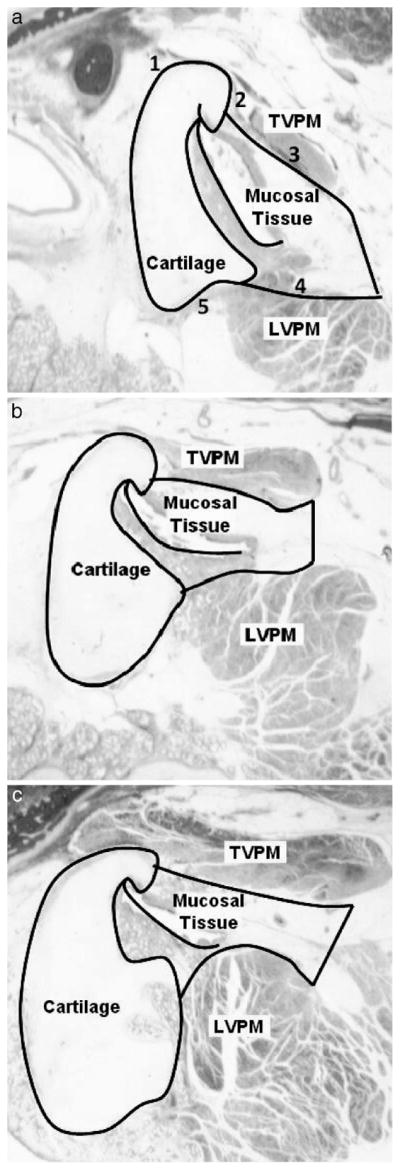
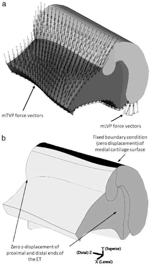
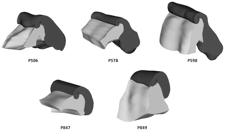
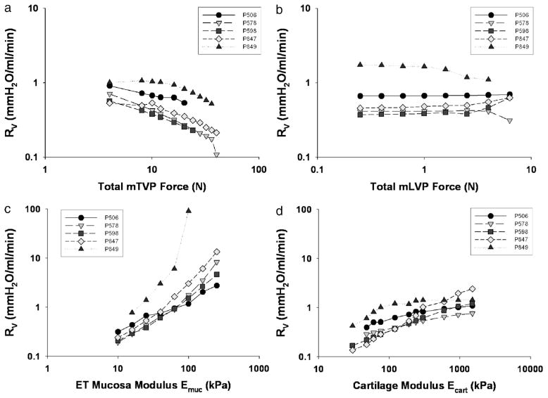
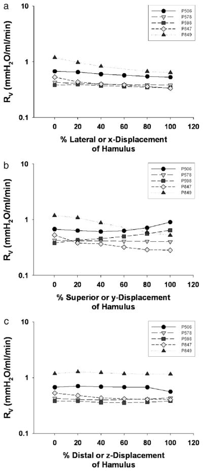
Methods: Finite element models of ET anatomy and physiology were developed by using image analysis and three-dimensional (3D) reconstruction techniques. Models were developed using histological images of ET structures obtained from five infant CP specimens. The models were parameterized, and the effects of varying model parameters, which included tensor veli palatini and levator veli palatini force, ET cartilage, periluminal mucosal compliance, and hamular position on resistance to airflow through the tubal lumen, were determined.
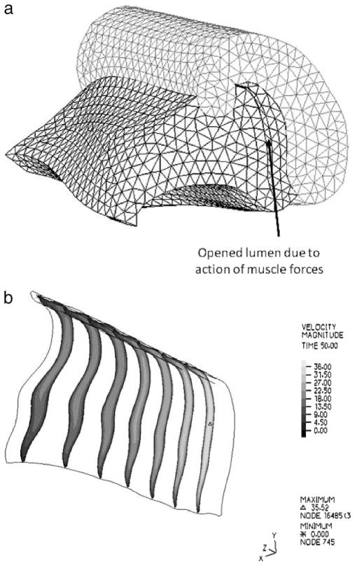
Results: Of the evaluated parameters, only applied tensor veli palatini muscle force and compliance of the periluminal mucosa and cartilage tissues were significant predictors of resistance to airflow through the ET during muscle-assisted opening.
Conclusions: Finite element models of ET function in the CP infant identified tensor veli palatini muscle force as a direct predictor and mucosal/cartilage compliance as an indirect predictor of ET opening during muscle-assisted lumen dilations. Hamular position and levator veli palatini force were not found to have an effect on ET function in CP infants.
Figures
Similar articles
-
A fresh cadaveric study of the paratubal muscles: implications for eustachian tube function in cleft palate.Plast Reconstr Surg. 1997 Sep;100(4):833-42. doi: 10.1097/00006534-199709001-00003. Plast Reconstr Surg. 1997. PMID: 9290650
-
The role of the tensor veli palatini muscle in the development of cleft palate-associated middle ear problems.Clin Oral Investig. 2016 Sep;20(7):1389-401. doi: 10.1007/s00784-016-1828-x. Epub 2016 May 7. Clin Oral Investig. 2016. PMID: 27153847 Free PMC article. Review.
-
Three-dimensional finite element analysis of Eustachian tube function under normal and pathological conditions.Med Eng Phys. 2012 Jun;34(5):605-16. doi: 10.1016/j.medengphy.2011.09.008. Epub 2011 Oct 12. Med Eng Phys. 2012. PMID: 21996354 Free PMC article.
-
Timing of tensor and levator veli palatini force application determines eustachian tube resistance patterns during the forced-response test.Auris Nasus Larynx. 2010 Dec;37(6):720-9. doi: 10.1016/j.anl.2010.02.008. Epub 2010 Apr 21. Auris Nasus Larynx. 2010. PMID: 20413236 Free PMC article.
-
Relevance of the pharyngotympanic tube.SADJ. 2003 Sep;58(8):335-7. SADJ. 2003. PMID: 14648916 Review.
Cited by
-
Quantification of nasal airflow resistance in English bulldogs using computed tomography and computational fluid dynamics.Vet Radiol Ultrasound. 2017 Sep;58(5):542-551. doi: 10.1111/vru.12531. Epub 2017 Jul 17. Vet Radiol Ultrasound. 2017. PMID: 28718208 Free PMC article.
-
Computational analysis of microbubble flows in bifurcating airways: role of gravity, inertia, and surface tension.J Biomech Eng. 2014 Oct;136(10):101007. doi: 10.1115/1.4028097. J Biomech Eng. 2014. PMID: 25068642 Free PMC article.
-
Imaging of the Eustachian tube and its function: a systematic review.Neuroradiology. 2016 Jun;58(6):543-556. doi: 10.1007/s00234-016-1663-4. Epub 2016 Feb 27. Neuroradiology. 2016. PMID: 26922743 Free PMC article.
-
The mechanism of balloon Eustachian tuboplasty: a biomechanical study.Med Biol Eng Comput. 2020 Apr;58(4):689-699. doi: 10.1007/s11517-020-02121-z. Epub 2020 Jan 17. Med Biol Eng Comput. 2020. PMID: 31953796 Free PMC article.
-
Multi-scale finite element modeling of Eustachian tube function: influence of mucosal adhesion.Int J Numer Method Biomed Eng. 2016 Dec;32(12):10.1002/cnm.2776. doi: 10.1002/cnm.2776. Epub 2016 Mar 22. Int J Numer Method Biomed Eng. 2016. PMID: 26891171 Free PMC article.
References
-
- Bathe KJ. Finite Element Procedures. Upper Saddle River, NJ: Prentice Hall; 1996.
-
- Bluestone CD. Eustachian tube obstruction in the infant with cleft palate. Ann Otol Rhinol Laryngol. 1971;80(suppl 2):1–30. - PubMed
-
- Bluestone CD, Beery QC, Cantekin EI, Paradise JL. Eustachian tube ventilatory function in relation to cleft palate. Ann Otol Rhinol Laryngol. 1975;84:333–338. - PubMed
-
- Bluestone CD, Doyle WJ. Anatomy and physiology of eustachian tube and middle ear related to otitis media. J Allergy Clin Immunol. 1988;81:997–1003. - PubMed
-
- Butow KW, Louw B, Hugo SR, Grimbeeck RJ. Tensor veli palatini muscle tension sling for Eustachian tube function in cleft palate: surgical technique and audiometric examination. J Craniomaxillofac Surg. 1991;19:71–76. - PubMed
Publication types
MeSH terms
Grants and funding
LinkOut - more resources
Full Text Sources
Medical
Miscellaneous
