. 2010 May 25:10:12.
doi: 10.1186/1472-6807-10-12.

Progesterone modulation of transmembrane helix-helix interactions between the alpha-subunit of Na/K-ATPase and phospholipid N-methyltransferase in the oocyte plasma membrane

Affiliations

Progesterone modulation of transmembrane helix-helix interactions between the alpha-subunit of Na/K-ATPase and phospholipid N-methyltransferase in the oocyte plasma membrane

Gene A Morrill et al. BMC Struct Biol. .

Abstract

Background: Progesterone binding to the surface of the amphibian oocyte initiates the meiotic divisions. Our previous studies with Rana pipiens oocytes indicate that progesterone binds to a plasma membrane site within the external loop between the M1 and M2 helices of the alpha-subunit of Na/K-ATPase, triggering a cascade of lipid second messengers and the release of the block at meiotic prophase. We have characterized this site, using a low affinity ouabain binding isoform of the alpha1-subunit.

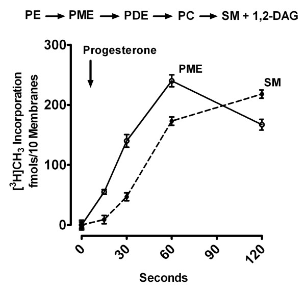
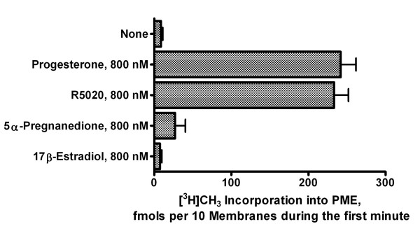
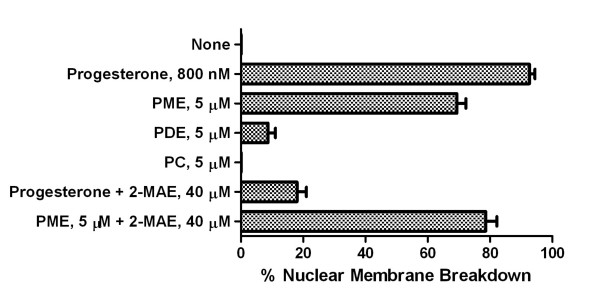
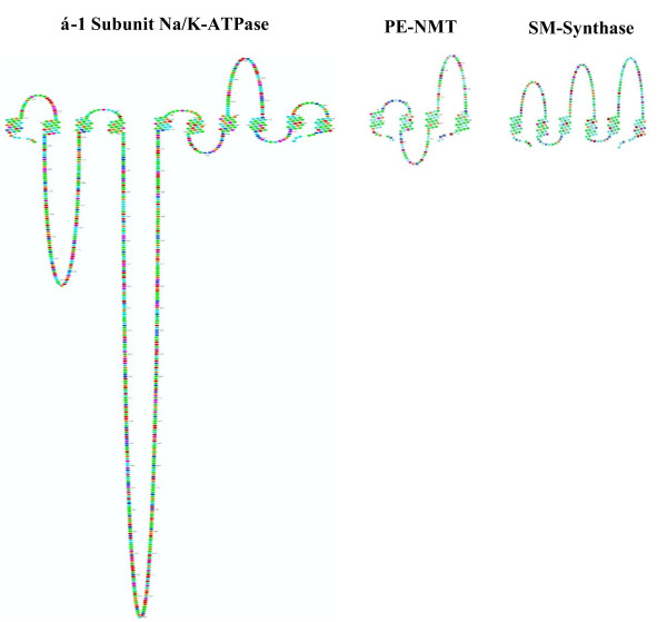
Results: Preparations of isolated plasma membranes from Rana oocytes demonstrate that physiological levels of progesterone (or the non-metabolizable progestin R5020) successively activate phosphatidylethanolamine-N-methyltransferase (PE-NMT) and sphingomyelin synthase within seconds. Inhibition of PE-NMT blocks the progesterone induction of meiosis in intact oocytes, whereas its initial product, phosphatidylmonomethylethanolamine (PME), can itself initiate meiosis in the presence of the inhibitor. Published X-ray crystallographic data on Na/K-ATPase, computer-generated 3D projections, heptad repeat analysis and hydrophobic cluster analysis of the transmembrane helices predict that hydrophobic residues L, V, V, I, F and Y of helix M2 of the alpha1-subunit interact with F, L, G, L, L and F, respectively, of helix M3 of PE-NMT.

Conclusion: We propose that progesterone binding to the first external loop of the alpha1-subunit facilitates specific helix-helix interactions between integral membrane proteins to up-regulate PE-NMT, and, that successive interactions between two or more integral plasma membrane proteins induce the signaling cascades which result in completion of the meiotic divisions.


Figures

Figure 1
Figure 1
Time course of [3H]Methyl incorporation into PME and SM. Upper: The steps involved in phosphatidylethanolamine (PE) N-methylation and sphingomyelin (SM) synthesis) in R. Pipiens oocyte plasma membranes involving PE, phosphatidylmonomethylethanolamine (PME), phosphatidyldimethylethanolamine (PDE), phosphatidylcholine (PC) and sphingomyelin (SM. Lower: Net increase in S-adenosyl methionine-derived 3H ([3H]SAM) incorporation into PME and SM in isolated plasma-vitelline membranes as a function of time after addition of progesterone. The values shown are calculated from 3H migrating with phospholipid standards using one-dimensional TLC and are expressed as fmols per 10 membranes corrected for basal levels of the individual phospholipids at the times points indicated. 1,2-DAG (1,2-diacylglycerol) is the product of SM synthase. Values are means ± SEM for oocytes from 3 females.
Figure 2
Figure 2
Induction of PME formation in isolated oocyte plasma membranes membrane by exogenous progesterone, R5020 (a non-metabolizable progestin), 5α-3,20-pregnanedione, and 17β-estradiol. Plasma-vitelline membranes isolated from denuded prophase-arrested R. pipiens oocytes were preincubated with [3H]S-adenosyl methionine ([3H]SAM) for 2 min at 20°C before addition of 800 nM (final concentration) of the steroid indicated. Individual samples containing 5-6 isolated membranes were frozen in liquid nitrogen at 0, 15, 30, 60 and 120 s after progesterone addition. Membranes were then extracted and analyzed for [3H]phosphatidylmonomethylethanolamine (PME) as described in Methods. Values are means ± SEM for oocytes from 3 females.
Figure 3
Figure 3
The induction of meiosis by progesterone compared to its induction by the products of phosphatidylethanolamine N-methylation. Denuded R. pipiens oocytes were transferred to Ringer's solution containing progesterone, PME, PDE and PC and incubated at 20-22°C. These phospholipids, when sonicated in Ringer's solution, formed clear solutions that were stable for several hours at room temperature. Denuded oocytes incubated in the phospholipid-Ringer's micelles for 6 h were rinsed, and then transferred to Ringer's solution for 6 h and nuclear membrane breakdown measured as described in methods. Sibling oocytes were preincubated in an N-methylation inhibitor [2-Methyl(amino)ethane (2-MAE)] for 1 h and transferred to Ringer's solution containing 2-MAE and progesterone or PME. Phospholipids were suspended in Ringer's solution as described in Methods. Results are expressed as means ± SEM for oocyte preparations from 3 females.
Figure 4
Figure 4
The topology of the α1-subunit of the Na/K-ATPase (left), PE-N-methyltransferase (center), and SM synthase (right) in Scalable Vector graphics (SVG) format [14]. The cross section of each enzyme is shown with the lumenal side uppermost and the intracellular environment at the bottom. The N and C termini of all three proteins are located intracellularly.
Figure 5
Figure 5
Architecture of the α1-subunit of the Na/K-ATPase αβγ complex. α-helices are represented by ribbons and β-strands by arrows with wires that follow the backbone of the α-carbon loops. The β subunit is indicated in blue and the γ subunit in green. The 3D conformation shown is based on the x-ray crystallographic coordinates [12] as analyzed using Pymol Molecular Viewer, ver. 1.1 (DeLano Scientific LLC (pymol.sourceforgr.net). The plasma membrane region is indicated by the red and blue dotted lines with the top as extracellular and bottom as intracellular.
Figure 6
Figure 6
The 10 transmembrane domains of the Na/K-ATPase α1-subunit seen from the cytoplasmic surface (upper cartoon) and as a transmembrane projection (lower cartoon). The x-ray crystallographic coordinates [12] were analyzed using Pymol Molecular Viewer, ver. 1.1 (DeLano Scientific LLC (pymol.sourceforgr.net) and King Display Software, ver. 2.14 (kinnemage.biochem.duke.edu/software/king.php) to generate the upper and upper cartoons, respectively. The relative positions of the lower helices are shown with a 12 ± 2°tilt angle.
Figure 7
Figure 7
Comparison of the 3D projections of the 10 transmembrane (M1 - M10) helices of the α1-subunit of the Na, K-ATPase. Projections are shown as if traversing the plasma membrane from extracellular (top) to intracellular (bottom). Plots were generated using Chem 3D Ultra v. 11.0 (Cambridgesoft.com). Colors indicate individual amino acids.
Figure 8
Figure 8
The 3D projection of the M2 transmembrane helix of the α1-subunit of Na/K-ATPase (center) compared to the corresponding hydrophobic cluster analysis (left) and its relationship to the lipid bilayer of the plasma membrane (right). The center projection illustrates the sequence L134 to A154 (Rat Primary accession # PO6685) written in the classical α-helix (3.6 amino acids per turn). The left projection indicates the hydrophobic cluster analysis [15] after unrolling the helix (see text) with the hydrophobic clusters circled. Filled diamonds indicate glycine, squares with a point (serine) or not (threonine), and C for cysteine. The right projection depicts a stylized crystal cartoon of a lipid bilayer containing phosphatidylcholine but no cholesterol. The cartoon is a RasMol image of a phosphatidylcholine bilayer published by E. Martz (see Methods). Green spheres represent water oxygens, blue represent lipid nitrogen, red represent lipid oxygen, and yellow represent lipid phosphorus.
Figure 9
Figure 9
The hydrophobic cluster analysis of M1 and M2 helices of the α1-subunit of the Na/K-ATPase (left column) compared with a comparable analysis of all four transmembrane helices (M1-M2-M3-M4) of PE-N-methyltransferase (PE-NMT) (right column). A red star indicates proline, filled diamonds indicate glycine, squares with a point (serine) or not (threonine), and C for cysteine. Each projection is displayed with the helix unrolled with the top facing the lunenal surface of the cell.
Figure 10
Figure 10
Helical wheel representations of the amino acid sequences of the M2 helix of the α1-subunit of the Na, K-ATPase and helix M3 of the PE NMT. The amino acid side chains are projected down the axis of the alpha helix (orthogonal to the plane of the page). The nonpolar residues are in yellow, the polar, uncharged residues are green. As an ideal alpha helix consists of 3.6 residues per complete turn, the angle between two residues is chosen to be 100 degrees and thus there exists a periodicity after five turns and 18 residues. The Figure is a snapshot of a Java Applet written by Edward K. O'Neil and Charles M. Grisham, University of Virginia at Charlottsville, VA (see Methods).

Similar articles

Cited by

References

    1. Morrill GA, Kostellow AB. Progesterone induces meiotic division in the amphibian oocyte by releasing lipid messengers from the plasma membrane. Steroids. 1999;64:157–67. doi: 10.1016/S0039-128X(98)00093-2. - DOI - PubMed
    1. Morrill GA, Erlichman J, Gutierrez-Juarez R, Kostellow AB. The steroid binding subunit of the Na/K-ATPase as a progesterone receptor on the amphibian oocyte plasma membrane. Steroids. 2005;70:933–945. doi: 10.1016/j.steroids.2005.07.002. - DOI - PubMed
    1. Morrill GA, Kostellow AB, Askari A. Progesterone binding to the alpha1-subunit of the Na/K-ATPase on the cell surface: Insights from computational modeling. Steroids. 2008;70:933–45. doi: 10.1016/j.steroids.2005.07.002. - DOI - PMC - PubMed
    1. Blostein R. Jeanne Mannery Fisher Memorial Lecture 1988. Structure-function studies of the sodium pump. Biochem Cell Biol. 1999;77:1–10. doi: 10.1139/bcb-77-1-1. - DOI - PubMed
    1. Blanco G, Mercer RW. Isozymes of the Na/K-ATPase: heterogeneity in structure, diversity in function. Am J Physiol. 1998;275(5 Pt 2):F633–50. - PubMed

Publication types

MeSH terms

Substances