. 2010 Aug 15;344(2):669-81.
doi: 10.1016/j.ydbio.2010.05.496. Epub 2010 May 23.

Mesodermal Tbx1 is required for patterning the proximal mandible in mice

Affiliations

Mesodermal Tbx1 is required for patterning the proximal mandible in mice

Vimla S Aggarwal et al. Dev Biol. .

Abstract

Defects in the lower jaw, or mandible, occur commonly either as isolated malformations or in association with genetic syndromes. Understanding its formation and genetic pathways required for shaping its structure in mammalian model organisms will shed light into the pathogenesis of malformations in humans. The lower jaw is derived from the mandibular process of the first pharyngeal arch (MdPA1) during embryogenesis. Integral to the development of the mandible is the signaling interplay between Fgf8 and Bmp4 in the rostral ectoderm and their downstream effector genes in the underlying neural crest derived mesenchyme. The non-neural crest MdPA1 core mesoderm is needed to form muscles of mastication, but its role in patterning the mandible is unknown. Here, we show that mesoderm specific deletion of Tbx1, a T-box transcription factor and gene for velo-cardio-facial/DiGeorge syndrome, results in defects in formation of the proximal mandible by shifting expression of Fgf8, Bmp4 and their downstream effector genes in mouse embryos at E10.5. This occurs without significant changes in cell proliferation or apoptosis at the same stage. Our results elucidate a new function for the non-neural crest core mesoderm and specifically, mesodermal Tbx1, in shaping the lower jaw.


Figures

FIGURE 1
FIGURE 1. Mandibular skeletal phenotype in Tbx1-/- embryos
(A-F) Skeletal preparations of E17.5 dpc embryos showing ossified areas in red and cartilage in blue. (A, B) Lateral view of head of wild-type Tbx1+/+ (A) and Tbx1-/- (B) embryo (see Fig. S1). (C, D) Mandible of a wild-type embryo (C) and a Tbx1-/- embryo (D). Cr, coronoid process; Cn, condylar process; Ang, angular process. (E, F) Ear capsule of Tbx1+/+ (E) and Tbx1-/- (G) embryo. Arrow denotes the tympanic ring in the Tbx1+/+ embryo (E), which is missing in the Tbx1-/- embryo (F). (G-L) Transverse histological sections of Tbx1+/+ (G, I, K) and Tbx1-/- embryo (H, J, L) stained with hematoxylin and eosin at E17.5. Incus and stapes are missing in the Tbx1-/- embryo (H) as compared with the Tbx1+/+ (G), see Fig. S2. s, stapes; i, incus; m, malleus. (I, J) Mandibular molar teeth are present in both the wild-type (I) and Tbx1 mutant embryos (J). mo, molar. (K, L) Mandibular incisors are present in both the wild-type (K) and Tbx1 mutant embryos (L). in, incisor. Tooth abnormalities were not detected at this level of magnification.
FIGURE 2
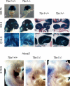
FIGURE 2. Localization of cranial neural crest cells in Tbx1-/-; Wnt1-Cre; R26R embryos
(A-F) Lateral views of Tbx1+/+; Wnt1-Cre; R26R (A, C, E) and Tbx1-/-; Wnt1-Cre; R26R (B, D, F) embryos. (A, B) At E8.75, β- gal stained cells (blue) are observed in both Tbx1+/+; Wnt1-Cre; R26R (A) and Tbx1-/-; Wnt1-Cre; R26R (B) extending from the neural tube in two streams towards pharyngeal arch one (1) and two (2), with no observed differences between the two embryos. (C, D) At E9.5, stained cells are seen in Tbx1+/+; Wnt1-Cre; R26R (C) and Tbx1-/-; Wnt1-Cre; R26R (D) in the frontonasal prominence, first (1) and second (2) pharyngeal arch. Arrow marks the additional stream of NCCs that migrate into the first pharyngeal arch from rhombomere 4 as compared with wild- type embryos. Second arch is missing from null mutants. (E, F) The staining pattern in E10.5 embryos is similar to that observed at E9.5, with stained cells present throughout the pharyngeal arches. The second and third arch are missing from the null mutants. (G-J) Histological analysis of NCCs in the pharyngeal arches of Tbx1-/-; Wnt1-Cre; R26R embryos. Tbx1+/+; Wnt1-Cre; R26R (G, I) and Tbx1-/-; Wnt1-Cre; R26R (H, J) embryos were stained in whole mount for β- galactosidase activity, paraffin embedded and sectioned. Most of the cells in the first pharyngeal arch are labeled except for cells in the centre of the arch that arise from the paraxial mesoderm. (K-M). Lateral views of whole mount in situ hybridization of embryos at E10.5 for Hoxa2 mRNA expression. Two Tbx1 -/- embryos are shown (L-M), one is more mild and has the second pharyngeal arch (L) and the other is more severe and is missing the second arch (M).
FIGURE 3
FIGURE 3. Wnt1-Cre; Tbx1flox/- embryos do not have a craniofacial phenotype
(A-N) Coronal sections of Wnt1- Cre; Tbx1flox/+ (controls: A, C, E, G, I, K and M) and Wnt1-Cre; Tbx1flox/- (NCC-KO; B, D, F, H, J, L and N) at E17.5. (A- B) There is no difference in the mandible (m) between the controls (A) and the mutant embryos (B). (C-D) Malleus (ma) is present in wildtype (C) and mutant embryos (D). (E-F) Tympanic ring (marked by arrow) forms in the controls (E) and mutant (F) embryos. (G-H) Mandibular molar teeth (mo) are present in the controls (G) and mutants (H). T, tongue. (I-J) Controls (I) as well as mutants (J) have mandibular incisors (i). (K-L) The palate is normal in the controls (K) and mutants (L). Arrow points to point of fusion of palatal shelves. (M-N) Masseter (mt), which is missing in the Tbx1-/- mice, is formed in both controls (M) and mutants (N).
FIGURE 4
FIGURE 4. Expression of neural crest mesenchymal transcription factors in the Tbx1-/- MdPA1 at E10.5
(A- H) are lateral views of the embryos; (A’- H’) are frontal views. (A, A’) In situ hybridization showing the distribution of Lhx6 transcripts in the mandibular arch of a Tbx1+/+ embryo. (B, B’) Lhx6 is expanded into the proximal and dorsal (proximal to the arch; dorsal to the embryo) region of the mandibular arch of Tbx1-/- embryos (arrows in B, B’). (C, C’) Lhx8 expression is seen in the anterior region of the mandibular arch in Tbx1+/+ embryos. (D, D’) Expression of Lhx8 is expanded in the proximo-dorsal region of the Tbx1-/- mandibular arch (arrows in D, D’). (E, E’) Ptx1 is expressed in a restricted domain of the mandibular arch of Tbx1+/+ embryos. (F, F’) The domain of expression of Ptx1 is changed in the Tbx1-/- embryo, with Ptx1 expanding in the proximo-dorsal region of the mandibular arch (arrows in F, F’). (G- H’) Gsc is expressed in the caudal mesenchyme of the mandibular arch of the Tbx1+/+ (G, G’) and Tbx1-/- (H, H’) embryos. Note subtle shape difference among embryos of similar genotypes (A’-H’). Right, cartoon depicting expression patterns in Tbx1+/+ and Tbx1-/- embryos. Arrow shows direction of shift to proximal side of MdPA1. Below, shows previously established model of genetic pathway.
FIGURE 5
FIGURE 5. Fgf signaling is altered in the mandibular arches of the Tbx1-/- embryos at E10.5
(A- H) are lateral views of the embryos; (A’- H’) are frontal views (A, A’) In situ hybridization showing the distribution of Spry1 transcripts in the mandibular arch of a Tbx1+/+ embryo. (B, B’) Spry1 is expanded into the proximal and posterior region of the mandibular arch of Tbx1-/- embryos (arrows in B, B’). (C, C’) Spry2 expression is seen in the anterior region of the mandibular arch in Tbx1+/+ embryos. (D, D’) Expression of Spry2 is expanded in the proximo-posterior region of the Tbx1-/- mandibular arch (arrows in D, D’). (E, E’) Erm is expressed in a restricted domain of the mandibular arch of Tbx1+/+ embryos. (F, F’) The domain of expression of Erm is changed in the Tbx1-/- embryo, with Erm expanding in the posterior and proximal region of the mandibular arch (arrows in F, F’). (G, G’) Fgf8 is expressed in the proximal oral ectoderm of the Tbx1+/+ embryos. (H, H’) Expression of Fgf8 is shifted in a proximo-caudal direction in the mandibular arches of the Tbx1-/- embryos (arrows in H, H’). Note varying MdPA1 shapes within genotypes. Right, cartoon depicting expression patterns in Tbx1+/+ and Tbx1-/- embryos. Arrow shows direction of shift to proximal side of MdPA1. Below, shows model of previously established genetic pathway.
FIGURE 6
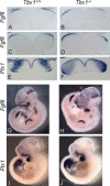
FIGURE 6. Expression of Fgf8 is altered in the Tbx1-/- MdPA1 at E10.5
(A- F) In situ hybridization on coronal sections of the Tbx1+/+ (A, C and E) and Tbx1-/- mutants (B, D and F) mandibular arches. (A, B) At E10.5, Fgf8 is expressed in the proximal oral ectoderm of the mandibular arch in the Tbx1+/+ embryo (A). Its expression is shifted in a proximal (lateral to the embryo) direction in the Tbx1-/- (B) embryos. (C, D) Similar expression changes are seen in the Tbx1-/- embryos (D) as compared with the Tbx1+/+ embryos (C) at E9.5. (E, F) Section in situ hybridization also shows the expansion of Ptx1 into the proximal region of the mandibular arch of Tbx1-/- embryo (F) compared with the wild-type (E). (G-J) Lateral views of whole mount in situ hybridization experiments for Fgf8 (G, H) and Ptx1 (I, J) in Tbx1+/+ (G, I) and Tbx1-/- embryos (H, J) embryos. Additional and frontal views are in Figs. 4 and 5.
FIGURE 7
FIGURE 7. Analysis of Bmp4 and downstream target genes in the Tbx1-/- MdPA1
(A-F, J-Q) Whole mount and (G-I; R-T) section in situ hybridization of Tbx1+/+ (A, C, E, G, J, L, N, P, R) and Tbx1-/- (B, D, F, H, I, K, M, O, Q, S, T) embryos at E10.5. (A, C, E, G) Bmp4 is expressed in the distal (midline) oral ectoderm of the mandibular arch of wild-type embryos. (B, D, F, H, I) Its expression is reduced but not absent in the distal midline and expanded into the proximal region of the Tbx1-/- mandibular arch. In situ hybridization on coronal sections shows the proximal expansion of Bmp4 in the Tbx1-/- mandibular arch (H, I) as compared with Tbx1+/+ (G). (J, L, N, P, R) Expression of Alx4, a Bmp4 target gene, is located under the Bmp4 expression domain in Tbx1+/+ embryos. (K, M, O, Q, S, T) Alx4 is shifted proximally in Tbx1-/- embryos (arrow in K) similar to the proximal shift in Bmp4 expression.
FIGURE 8
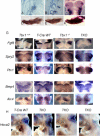
FIGURE 8. Mesodermal Tbx1 regulates mandibular arch patterning
(A- B) In situ hybridization of Tbx1 on sagittal sections at E8.5 (A), E9.5 (B) and a coronal whole mount view at E10.5 (C) show that Tbx1 is not expressed in the oral ectoderm of the mandibular arch at the stages examined. (D-F) Skeletal preparations of E17.5 embryos showing the lateral view of a mandible of T- Cre; Tbx1flox/+ (D), T- Cre; Tbx1flox/- (E) and Tbx1-/- (F) embryos. Phenotype is identical to Tbx1-/-, however penetrance is reduced. (F) Coronal views of whole mount in situ hybridization of E10.5, Tbx1+/+, Tbx1-/-, T- Cre; Tbx1flox/+ (T-Cre WT) and T- Cre; Tbx1flox/- (TKO) embryos using probes for Fgf8, Spry, Ptx1, Bmp4 and Alx4. Genes show a similar proximal shift in expression as in Tbx1-/- mutant embryos at E10.5. (H) Whole mount views of Hoxa2 whole mount in situ hybridization at E10.5. One three TKO mutants are shown with different amounts of reduction of the second pharyngeal arch, with the left being the most mild and the right being the most severe.
FIGURE 9
FIGURE 9. Shh expression is unaltered but Ptch1 is missing from the distal midline downstream of Tbx1 in MdPA1 at E10.5
(A- D) Coronal tissue sections from Tbx1+/+ and Tbx1-/- embryos probed to detect Shh mRNA, shows similar expression patterns in the ventral neural tube and ectoderm of the first pharyngeal arch. (E-H) Coronal views of whole mount in situ hybridization of E10.5 embryos for Ptch1, a downstream gene of Shh. Ptch1 expression is present in the distal midline and proximal sides of MdPA1 in Tbx1+/+ embryos (E, G) but expression was reduced or missing from the distal midline in Tbx1-/- mutant embryos (F, H). Below is a cartoon illustration of the changes observed (Ptch1 = purple).
FIGURE 10
FIGURE 10. No difference in proliferation or apoptosis between Tbx1+/+ and Tbx1-/- embryos at E10.5
(A,B) Sections through location C on illustration of first arch (center). (A) At E10.5, proliferation is seen more concentrated in the yellow zones of the first arch. (B) Apoptosis is located very specifically through the medial (distal) portion of the first arch, depicted as the red zone (image). (A’, B’) Zones of proliferation and apoptosis are highlighted and enlarged below. (Table) Results from counting cells stained for proliferation and apoptosis are shown. Statistical analysis by T-test confirms no statistical significance in the difference between cells stained for proliferation or apoptosis in Tbx1+/+ and Tbx1-/- embryos. P value was accepted at p=0.05.
FIGURE 11
FIGURE 11. Model of Tbx1 function in MdPA1 patterning
Cartoon shows location of Tbx1 expression in the proximal core mesoderm (green) in Tbx1+/+ and Tbx1-/- embryos and E10.5. Below are expression domains of Fgf8 and Bmp4. Model shows Tbx1 restricting proximal expression domains of Fgf8 and Bmp4. Genetic pathway for establishing axes forming the proximal lower jaw downstream of Tbx1. Genes expressed in the different germ layers comprising MdPA1 are indicated (endoderm, black; ectoderm, blue; mesoderm, green; neural crest cells, red). References for the pathway are provided in the text.

References

    1. Ahlgren SC, Bronner-Fraser M. Inhibition of sonic hedgehog signaling in vivo results in craniofacial neural crest cell death. Curr Biol. 1999;9:1304–1314. - PubMed
    1. Alappat SR, Zhang Z, Suzuki K, Zhang X, Liu H, Jiang R, Yamada G, Chen Y. The cellular and molecular etiology of the cleft secondary palate in Fgf10 mutant mice. Dev Biol. 2005;277:102–113. - PubMed
    1. Arnold JS, Werling U, Braunstein EM, Liao J, Nowotschin S, Edelmann W, Hebert JM, Morrow BE. Inactivation of Tbx1 in the pharyngeal endoderm results in 22q11DS malformations. Development. 2006;133:977–987. - PubMed
    1. Beresford WA. Schemes of zonation in the mandibular condyle. Am J Orthod. 1975;68:189–195. - PubMed
    1. Beverdam A, Brouwer A, Reijnen M, Korving J, Meijlink F. Severe nasal clefting and abnormal embryonic apoptosis in Alx3/Alx4 double mutant mice. Development. 2001;128:3975–3986. - PubMed

Publication types

MeSH terms