. 2010 Jun;137(12):2045-54.
doi: 10.1242/dev.043299.

FOXA1 is an essential determinant of ERalpha expression and mammary ductal morphogenesis

Affiliations

FOXA1 is an essential determinant of ERalpha expression and mammary ductal morphogenesis

Gina M Bernardo et al. Development. 2010 Jun.

Abstract

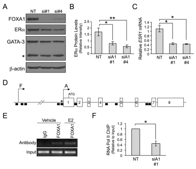
FOXA1, estrogen receptor alpha (ERalpha) and GATA3 independently predict favorable outcome in breast cancer patients, and their expression correlates with a differentiated, luminal tumor subtype. As transcription factors, each functions in the morphogenesis of various organs, with ERalpha and GATA3 being established regulators of mammary gland development. Interdependency between these three factors in breast cancer and normal mammary development has been suggested, but the specific role for FOXA1 is not known. Herein, we report that Foxa1 deficiency causes a defect in hormone-induced mammary ductal invasion associated with a loss of terminal end bud formation and ERalpha expression. By contrast, Foxa1 null glands maintain GATA3 expression. Unlike ERalpha and GATA3 deficiency, Foxa1 null glands form milk-producing alveoli, indicating that the defect is restricted to expansion of the ductal epithelium, further emphasizing the novel role for FOXA1 in mammary morphogenesis. Using breast cancer cell lines, we also demonstrate that FOXA1 regulates ERalpha expression, but not GATA3. These data reveal that FOXA1 is necessary for hormonal responsiveness in the developing mammary gland and ERalpha-positive breast cancers, at least in part, through its control of ERalpha expression.


Figures

Fig. 1.
Fig. 1.
FOXA1 is expressed in the developing mammary gland in conjunction with ERα. (A) Representative images of FOXA1 and ERα IHC in virgin terminal end buds (TEBs) (5 weeks), virgin ductal epithelium (8 weeks), virgin alveoli (20 weeks), pregnant alveoli (day 18), lactating alveoli (day 2) and an involuting gland (day 5). Within the TEB, arrows mark the luminal progenitor cells and arrowheads mark the basal/myoepithelial progenitors. Unfilled arrowheads indicate expressing cells in the pregnant alveoli and involuting gland. FOXA1 and ERα expression (brown nuclei) is counterstained with Hematoxylin. (B) Representative image of dual IF for FOXA1 and ERα in virgin ductal epithelium (n=4). The luminal epithelium consists of four populations: cells co-expressing FOXA1 and ERα (31.8±4.4%) (yellow cells in ‘Merge’), expressing FOXA1 (12.1±5.0%) or ERα (3.8±0.6%) alone (arrowheads), or expressing neither (52.3±6.8%). Scale bars: 20 μm.
Fig. 2.
Fig. 2.
FOXA1 is required for mammary ductal outgrowth in an orthotopic transplantation model. (A-C) Representative whole mounts of ductal outgrowths arising from mammary anlagen collected from E14 Foxa1+/+ and Foxa1−/− mice and transplanted into cleared fat pads of 3- to 4-week-old syngeneic C57BL/6 recipients. (A) Recipients aged 5 weeks post-transplant. (B) Recipients aged 8 weeks post-transplant. (C) Recipients aged 8 weeks post-transplant with subsequent pregnancy (18.5 dpc). Epidermal cysts form as a result of co-transplantation of hair follicles along with the mammary gland. The number and percentage of mammary outgrowths for each donor genotype is indicated. Scale bars: 2 mm. *, epidermal cysts.
Fig. 3.
Fig. 3.
FOXA1 is required for TEB formation and ductal invasion. (A,B) Representative whole mounts of renal grafts of Foxa1+/+ and Foxa1−/− mammary glands (into wild-type C57BL/6 recipients) harvested at (A) 2 weeks (+/+, n=4; −/−, n=3) and (B) 4-5 weeks post-transplantation (+/+, n=4; −/−, n=4). Broken lines outline the mammary fat pad. (C) Quantitative real-time PCR of Foxa1 mRNA levels in the MaSC-enriched population (CD24+/CD29hi), the luminal progenitor population (CD24+/CD29lo/CD61+), and the mature luminal population (CD24+/CD29lo/CD61-) isolated from wild-type FVB/N inguinal mammary glands (n=10 per independent experiment). The results of two independent cell-sorting experiments are shown. Values were normalized to 18S rRNA (Exp#1) or Gapdh mRNA (Exp#2) and then expressed relative to the values obtained with the mature luminal population. Scale bars: 1 mm.
Fig. 4.
Fig. 4.
FOXA1 is not required for alveolar differentiation during pregnancy. (A) Representative whole mounts and Hematoxylin and Eosin-stained sections (+/+, n=5; −/−, n=3) and (B) images of milk protein IHC (brown) (+/+, n=3; −/−, n=3) in renal grafts from Foxa1+/+ and Foxa1−/− mammary glands harvested 4-5 weeks after transplantation and during late pregnancy (18.5 dpc). Sections were counterstained with Hematoxylin. Scale bars: 0.5 mm in A; 20 μm in B.
Fig. 5.
Fig. 5.
FOXA1 is required for expression of ERα in the normal mammary gland. (A) Representative images of ERα and PR IHC (brown nuclei) in renal grafts from Foxa1+/+ and Foxa1−/− mammary glands harvested 4-5 weeks post-transplantation (+/+, n=3; −/−, n=3). ERα and PR are maintained in the stroma of Foxa1−/− glands (arrows). (B) Foxa1, Pgr and Gata3 mRNA levels in renal transplanted Foxa1+/+ and Foxa1−/− mammary glands. Values represent the average ± s.d. and are relative to Krt8 mRNA (+/+, n=3; −/−, n=3; *P<0.01). (C) Quantitation of Foxa1 mRNA and (D) representative images of FOXA1 IHC (brown) in wild-type and Ex3αERKO mammary glands. Values represent the average ± s.d. and are relative to Krt8 mRNA (wild-type, n=3; Ex3αERKO, n=3). (E) Gata3 mRNA levels in wild-type and Ex3αERKO mammary glands. Values represent the average ± s.d. and are relative to Krt8 mRNA (wild-type, n=3; Ex3αERKO, n=3). (F) Representative images of FOXA1 IHC in Gata3+/f and MMTV-cre;Gata3f/f mammary glands (+/f, n=3; f/f, n=3). IHC quantification is depicted in the bottom right corner of each image. All sections were counterstained with Hematoxylin. Scale bars: 20 μm. NS, not significant.
Fig. 6.
Fig. 6.
FOXA1 regulates transcription of ESR1. (A-C) MCF7 cells were transiently transfected with non-targeting or two different siRNAs targeting FOXA1 (si#1 and si#4). (A) Representative immunoblots of FOXA1, ERα and GATA3 (I=3) [*, mutant form of GATA3 (Usary et al., 2004)]. (B) Quantitation of ERα protein levels relative to β-actin. Bars represent the mean of three experiments ± s.d. (*P<0.01; **P<0.005). (C) Quantitation of ESR1 mRNA levels. Bars represent the mean of three experiments ± s.d. relative to GAPDH mRNA (*P<0.005). (D) ESR1 is comprised of eight exons and at least seven promoters (only A and F are shown) (Reid et al., 2002). Regions previously identified to bind FOXA1 by ChIP-chip are indicated by black boxes (Lupien et al., 2008). (E) Representative (n=3) FOXA1 ChIP of the ESR1 promoter using primers amplifying a predicted binding site (* in D). MCF7 cells were treated with and without 17β-estradiol (E2). (F) MCF7 cells were transiently transfected with NT or FOXA1 si#1. Quantification of RNA polymerase II ChIP of the ESR1 promoter (n=3). Bars represent the average fold change relative to input and normalized relative to NT ± s.d. (*P<0.0005). NT, non-targeting siRNA.
Fig. 7.
Fig. 7.
Schematic of the mammary epithelial cell hierarchy. FOXA1 is expressed in and required for ductal development. GATA3 is expressed in and required for both ductal and alveolar development and is independent of FOXA1 expression. ERα is required for ductal and alveolar development, but is only expressed in ductal cells. This supports intercellular communication and/or lineage progression from ERα-positive ductal to ERα-negative alveolar cells (broken arrow).

References

    1. Abramoff M. D., Magelhaes P. J., Ram S. J. (2004). Image Processing with ImageJ. Biophotonics International 11, 36-42
    1. Asselin-Labat M. L., Sutherland K. D., Barker H., Thomas R., Shackleton M., Forrest N. C., Hartley L., Robb L., Grosveld F. G., van der Wees J., et al. (2007). Gata-3 is an essential regulator of mammary-gland morphogenesis and luminal-cell differentiation. Nat. Cell Biol. 9, 201-209 - PubMed
    1. Badve S., Turbin D., Thorat M. A., Morimiya A., Nielsen T. O., Perou C. M., Dunn S., Huntsman D. G., Nakshatri H. (2007). FOXA1 expression in breast cancer-correlation with luminal subtype A and survival. Clin. Cancer Res. 13, 4415-4421 - PubMed
    1. Behr R., Brestelli J., Fulmer J. T., Miyawaki N., Kleyman T. R., Kaestner K. H. (2004). Mild nephrogenic diabetes insipidus caused by Foxa1 deficiency. J. Biol. Chem 279, 41936-41941 - PubMed
    1. Besnard V., Wert S. E., Kaestner K. H., Whitsett J. A. (2005). Stage-specific regulation of respiratory epithelial cell differentiation by Foxa1. Am. J. Physiol. Lung Cell Mol. Physiol. 289, L750-L759 - PubMed

Publication types

MeSH terms

Substances