. 2010 Jul;24(7):1393-403.
doi: 10.1210/me.2010-0006. Epub 2010 May 25.

Granulosa cell-specific androgen receptors are critical regulators of ovarian development and function

Affiliations

Granulosa cell-specific androgen receptors are critical regulators of ovarian development and function

Aritro Sen et al. Mol Endocrinol. 2010 Jul.

Abstract

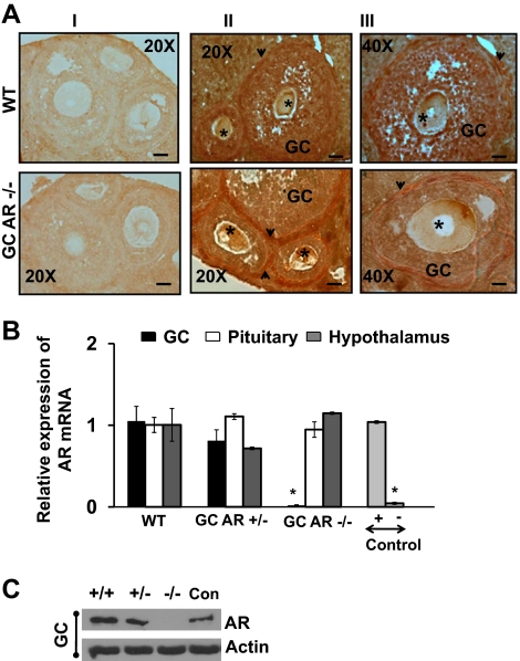
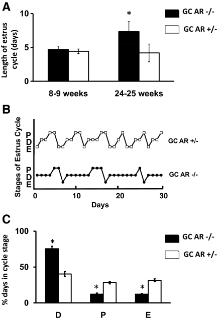
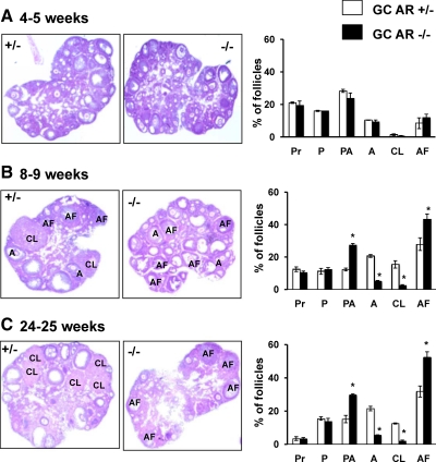
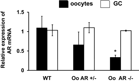
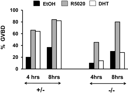
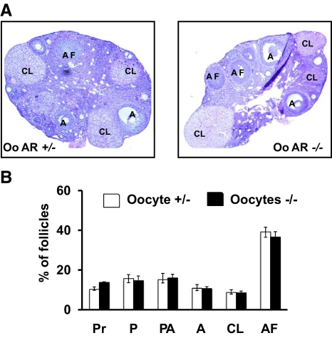
The physiological significance of androgens in female reproduction was unclear until female mice with global knockout of androgen receptor (AR) expression were found to have reduced fertility with abnormal ovarian function. However, because ARs are expressed in a myriad of reproductive tissues, including the hypothalamus, pituitary, and various ovarian cells, the role of tissue-specific ARs in regulating female fertility remained unknown. To examine the importance of ovarian ARs in female reproduction, we generated granulosa cell (GC)- and oocyte-specific AR-knockout (ARKO) mice by crossing AR-flox mice with MisRIIcre (GC-specific) or growth differentiation factor growth differentiation factor-9cre (oocyte-specific) mice. Relative to heterozygous and wild-type mice, GC-specific ARKO mice had premature ovarian failure and were subfertile, with longer estrous cycles and fewer ovulated oocytes. In addition, ovaries from GC-specific knockout mice contained more preantral and atretic follicles, with fewer antral follicles and corpus lutea. Finally, in vitro growth of follicles from GC-specific AR-null mice was slower than follicles from wild-type animals. In contrast to GC-specific AR-null mice, fertility, estrous cycles, and ovarian morphology of oocyte-specific ARKO mice were normal, although androgens no longer promoted oocyte maturation in these animals. Together, our data indicate that nearly all reproductive phenotypes observed in global ARKO mice can be explained by the lack of AR expression in GCs. These GC-specific ARs appear to promote preantral follicle growth and prevent follicular atresia; thus they are essential for normal follicular development and fertility.


Figures

Figure 1
Figure 1
Schematic diagram demonstrating the generation of GC- and oocyte-specific ARKO mouse. AR-loxPflox/flox females were crossed with GDF9cre males (for oocyte-specific −/−) or MisRIIcre males (for GC-specific −/−) and thereafter, the GDF9Cre/+ or MisRIICre/+;AR-loxPflox/+ females were backcrossed with AR-loxPflox/Y males to generate female heterozygous control GDF9- or MisRII-Cre/+;AR-loxPflox/+ mice and GDF9- or MisRII-Cre/+;AR-loxPflox/flox mice conditionally deleted for AR in the oocyte and GC, respectively.
Figure 2
Figure 2
Selective knockout of ARs in GCs. A, Representative photos of immunohistochemistry using an anti-AR antibody to detect AR expression in follicles isolated from 8- to 9-wk-old WT and GC-specific AR −/− mouse. The arrowhead and the star demonstrate AR theca cells and oocytes, respectively. Panel I represents the negative control (without primary antibody), and panels II and III represent follicles from WT and GC-specific AR −/− animals at ×20 (bar, 50 μm) and ×40 (bar, 100 μm) magnification, respectively. B, Graphic representation of AR mRNA transcripts in GCs, pituitary, and hypothalamus RNA extracts isolated from 8- to 9-wk-old WT, GC-specific AR +/−, and −/− animals. LnCAP and human embryonic kidney 293 cells were used as positive (+) and negative (−) controls, respectively. The mRNA levels were measured by quantitative real-time PCR and compared with control GAPDH mRNA expression using the ΔΔCt method. Results are represented as the amount relative to WT cells (mean ± se, n = 4 mice per genotype). *, Using ANOVA, P ≤ 0.001 for GC AR −/− vs. WT and GC AR +/− mice. C, Representative Western blot demonstrating the AR expression in GCs isolated from WT, GC-specific AR +/−, and −/− mice (n = 2 animals per genotype). LnCAP cells were used as positive control (Con). Similar results were observed in two separate experiments.
Figure 3
Figure 3
Knockout of the AR in GCs disrupt estrous cyclicity. A, Graphic representation of average length of estrous cycle determined by vaginal smears taken daily over a period of 3–4 wk in 8- to 9-wk- or 24- to 25-wk-old GC-specific AR −/− and +/− animals. B, Representative estrous cycles in a single GC-specific AR +/− (upper) and AR−/− (lower) mice. C, Percent of days spent in each estrous cycle stage (D, diestrus; P, proestrus; E, estrus) in GC-specific AR +/− and −/− mice. Data are represented as mean ± se (n = 5 mice). *, Using Student’s t test, P ≤ 0.05 for GC AR −/− vs. GC AR +/− mice.
Figure 4
Figure 4
GC-specific knockout of the AR results in reduced ovulation. A, Representative pictures of a cumulus-oocyte-complex (COC) and a denuded oocyte harvested from the oviduct and ampulla of a mouse after ovulation on the day of estrus (bar, 20 μm). B, Graphic representation of number of naturally ovulated oocytes in GC-specific AR −/− and AR +/− mice. Estrous cycles of 8- to 9-wk- and 24- to 25-wk-old female animals (n = 5 per genotype) were determined by vaginal smears for 2 wk and thereafter animals were killed at estrus. C, Number of superovulated oocytes in 8- to 9-wk- or 24- to 25-wk-old female GC-specific AR −/− and AR +/− mice (n = 5 per genotype). Mice were given a single ip injection of 5 U of pregnant mare serum gonadotropin followed 48 h later by 5 U of human chronic gonadotropin (Sigma). After 18 h, oocyte/cumulus masses were surgically isolated from the oviduct and ampulla and counted. Data are represented as mean ± se (n = 5 mice). *, Using Student’s t test P ≤ 0.01 for GC AR −/− vs. GC AR +/− mice.
Figure 5
Figure 5
GC-specific AR −/− mice have reduced follicle progression and increased follicle atresia relative to AR −/− females. Representative hematoxylin and eosin-stained ovarian sections and statistical analysis of percentage of different types of follicles from 4- to 5-wk- (A), 8- to 9-wk- (B), and 24- to 25-wk-old (C) GC-specific AR +/− and AR −/− mice (n = 4 ovaries per genotype). Sections were taken at intervals of 30 μm, and 5 μm paraffin-embedded sections were mounted on slides. The types of follicles counted are: primordial (Pr), primary (P), preantral (PA), antral (A), corpus luteum (CL), and atretic follicles (AF). *, Using Student’s t test, P ≤ 0.05 for GC AR −/− vs. AR +/− mice.
Figure 6
Figure 6
Ovarian follicles from GC-specific AR −/− mice grow more slowly in vitro than ovarian follicles from WT mice. Follicles of 100–120 μm diameter size were isolated from 21-d-old WT and GC AR −/− and cultured for 4 d in DMEM supplemented with 5 μg/ml of insulin, 5 μg/ml of transferrin, 5 ng/ml of sodium selenite, 1% penicillin-streptomycin, and 1 ng/ml of recombinant human FSH. Follicle diameters at the beginning (d0) and end (d4) of culture were measured by using the morphometric tool of the Zeiss Axioplan microscope (Zeiss). Representative pictures of follicles (panel A) and average diameter of follicles (panel B) (n = 25 follicles per genotype) isolated from GC-specific AR −/− and WT mice on d 0 and d 4 of culture. *, Using Student’s t test, P ≤ 0.05 for GC AR −/− vs. WT mice. Bar, 50 μm.
Figure 7
Figure 7
Selective knockout of the AR in oocytes (Oo). Relative expression of AR mRNA in the oocytes and GCs isolated from 8- to 9-wk-old WT and oocyte-specific AR −/− and +/− mice. mRNA levels were measured by quantitative real-time PCR and compared with control GAPDH mRNA expression using the ΔΔCt method. Results are represented as mean ± se (n = 4 per genotype). Approximately ≅120 oocytes per animal per genotype for each PCR were used. *, Using ANOVA, P ≤ 0.01 for GC AR −/− vs. WT and GC AT +/− mice.
Figure 8
Figure 8
Oocytes require AR expression for androgen- but not progestin-mediated maturation. Denuded oocytes isolated from 21-d-old oocyte-specific AR −/− and +/− mice (n = 2 animals per genotype) were treated with DHT (100 nm), R5020 (Promegestone, 250 nm), or ethanol (0.1%). Maturation (GVBD) of denuded oocytes was scored at 4 h and 8 h after treatment. Twenty-five oocytes per animal for each treatment were used for the experiment, and data are represented as the combined percent of oocytes that had undergone GVBD from two separate experiments. Data were analyzed using χ2 test and P ≤ 0.01 for DHT-treated oocytes isolated from oocyte-specific AR −/− vs. +/− mice.
Figure 9
Figure 9
Ovaries from oocyte (Oo)-specific AR +/− and AR −/− females are nearly identical. Representative hematoxylin and eosin-stained ovarian sections (upper panel) and statistical analysis of percentage of different type of follicles (lower panel) from 8- to 9-wk-old oocyte-specific AR +/− and −/− mice (n = 4 ovaries per genotype). Sections were taken at intervals of 30 μm and 5 μm paraffin-embedded sections were mounted on slides. The types of follicles counted are: primordial (Pr), primary (P), preantral (PA), antral (A), corpus luteum (CL), and atretic follicles (AF).

Similar articles

Cited by

References

    1. Quigley CA, De Bellis A, Marschke KB, el-Awady MK, Wilson EM, French FS 1995 Androgen receptor defects: historical, clinical, and molecular perspectives. Endocr Rev 16:271–321 - PubMed
    1. Lange CA, Gioeli D, Hammes SR, Marker PC 2007 Integration of rapid signaling events with steroid hormone receptor action in breast and prostate cancer. Annu Rev Physiol 69:171–199 - PubMed
    1. Xu Y, Chen SY, Ross KN, Balk SP 2006 Androgens induce prostate cancer cell proliferation through mammalian target of rapamycin activation and post-transcriptional increases in cyclin D proteins. Cancer Res 66:7783–7792 - PubMed
    1. Hillier SG, Whitelaw PF, Smyth CD 1994 Follicular oestrogen synthesis: the ‘two-cell, two-gonadotrophin’ model revisited. Mol Cell Endocrinol 100:51–54 - PubMed
    1. Simpson ER 2002 Aromatization of androgens in women: current concepts and findings. Fertil Steril 77(Suppl 4):S6–S10 - PubMed

Publication types

MeSH terms

Substances