. 2010 Jul;24(7):1338-48.
doi: 10.1210/me.2009-0402. Epub 2010 May 25.

Androgen receptor counteracts Doxorubicin-induced cardiotoxicity in male mice

Affiliations

Androgen receptor counteracts Doxorubicin-induced cardiotoxicity in male mice

Yasumasa Ikeda et al. Mol Endocrinol. 2010 Jul.

Abstract

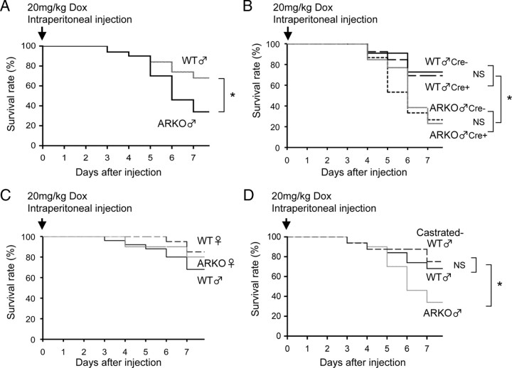
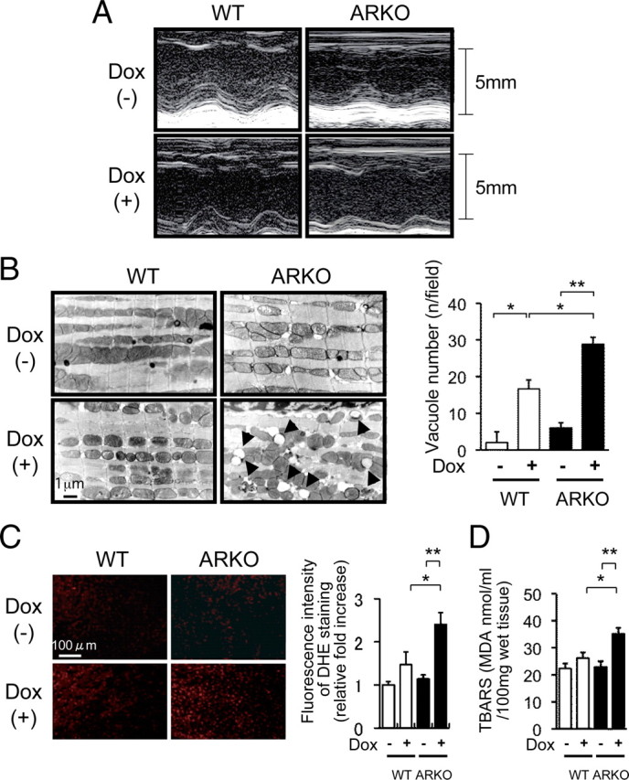
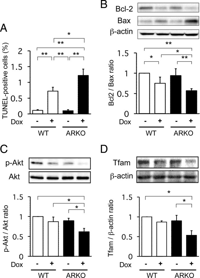
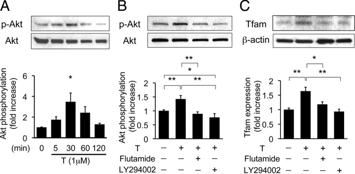
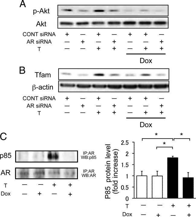
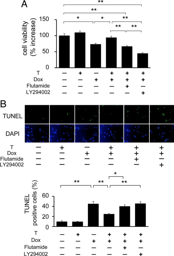
Doxorubicin (Dox) has been used as a potent anticancer agent, but serious cardiotoxicity precludes its use in a wide range of patients. We have reported that the androgen-androgen receptor (AR) system plays important roles in cardiac growth and protection from angiotensin II-induced cardiac remodeling. The present study was undertaken to clarify whether the androgen-AR system exerts a cardioprotective effect against Dox-induced cardiotoxicity. Male AR knockout (ARKO) and age-matched littermate male wild-type (WT) mice at 25 wk of age were given ip injections of Dox (20 mg/kg) or a vehicle. The survival rate and left ventricular function in Dox-treated male ARKO mice were reduced compared with those in Dox-treated male WT mice. Electron microscopic study showed prominent vacuole formation of myocardial mitochondria in Dox-treated male ARKO mice. Cardiac oxidative stress and apoptosis of cardiomyocytes were increased more prominently by Dox treatment in male ARKO mice than in male WT mice. In addition, Dox-induced reduction in the expression of cardiac mitochondria transcription factor A (Tfam) and phosphorylation of serine-threonine kinase (Akt) was more pronounced in male ARKO mice than in male WT mice. In cardiac myoblast cells, testosterone up-regulated Akt phosphorylation and Tfam expression and exerted an antiapoptotic effect against Dox-induced cardiotoxicity. Collectively, the results demonstrate that Dox-induced cardiotoxicity is aggravated in male ARKO mice via exacerbation of mitochondrial damage and superoxide generation, leading to enhanced apoptosis of cardiomyocytes. Thus, the androgen-AR system is thought to counteract Dox-induced cardiotoxicity partly through activation of the Akt pathway and up-regulation of Tfam to protect cardiomyocytes from mitochondrial damage and apoptosis.


Figures

Fig. 1.
Fig. 1.
Survival rates of male and female WT mice, male and female ARKO mice, and castrated male WT mice after Dox administration. Survival rates were calculated by the Kaplan-Meier method and compared by the log-lank test. n = 50 in each group. *, P < 0.05. NS, Not significant.
Fig. 2.
Fig. 2.
Left ventricular function, electron microscopic findings, superoxide generation, and lipid peroxidation after Dox administration in male WT mice and male ARKO mice. A, Representative M-mode echocardiogram of left ventricular wall motion. M-mode echocardiogram of the left ventriculum at 5 d after administration of the vehicle or Dox in male WT and male ARKO mice. Although there were slight extensional changes in left ventricular end-diastolic dimension (LVDd) and left ventricular end-systolic dimension (LVDs) of Dox-treated male WT mice, acute dilatation of LVDd and LVDs was markedly apparent in Dox-treated male ARKO mice. B, left panel, Electron microscopic findings of the left ventricular myocardium after administration of the vehicle or Dox in male WT and male ARKO mice. Accelerated vacuole formation of myocardial mitochondria was observed in Dox-treated male ARKO mice. Right panel, Quantitative analysis of vacuole number after administration of the vehicle or Dox in male WT (white bars) and male ARKO (black bars) mice. Values are expressed as means ± sem. *, P < 0.05; **, P < 0.01, n = 4–6 in each group. C, DHE bromide staining analysis after administration of the vehicle or Dox in male WT and male ARKO mice. D, Myocardial TBARS assay after administration of the vehicle or Dox in male WT (white bars) and male ARKO (black bars) mice. Values are expressed as means ± sem. *, P < 0.05; **, P < 0.01, n = 6–8 in each group.
Fig. 3.
Fig. 3.
Apoptosis of cardiac cells, cardiac Akt activation, and Tfam expression after Dox treatment in male WT and male ARKO mice. A, Numbers of TUNEL-positive cells in three separate left ventricular sections per mouse after administration of the vehicle or Dox in male WT (white bars) and male ARKO (black bars) mice. Values are expressed as means ± sem. *, P < 0.05; **, P < 0.01, n = 6 in each group. B, Cardiac expression of Bcl-2 and Bax. Upper panel, Representative blots of Bcl-2, Bax, and β-actin. Lower panel, Results of densitometric analysis of Bcl-2-to-Bax ratio after administration of the vehicle or Dox in male WT (white bars) and male ARKO (black bars) mice. Values are expressed as means ± sem. *, P < 0.05, n = 8 in each group. C, Cardiac Akt phosphorylation in male WT mice and male ARKO mice with or without Dox. Upper panels, Representative blots of phosphorylated forms of Akt and total Akt. Lower panels, Results of densitometric analysis of phosphorylated Akt (p-Akt) after administration of the vehicle or Dox in male WT (white bars) and male ARKO (black bars) mice. Values are expressed as means ± sem. *, P < 0.05, n = 8 in each group. D, Cardiac expression of Tfam with or without Dox treatment. Right panel, Representative blots of Tfam protein with results of densitometric analysis of Tfam expression after administration of the vehicle or Dox in male WT (white bars) and male ARKO (black bars) mice. Values are expressed as means ± sem. *, P < 0.05, n = 6 in each group.
Fig. 4.
Fig. 4.
Acute effect of androgen action on Akt phosphorylation (p-Akt) and Tfam expression in H9c2 cells. A, upper panel, Representative blots of phosphorylated Akt and total Akt. Lower panel, Results of densitometric analysis of phosphorylated Akt. Values are expressed as means ± sem. *, P < 0.05 vs. with control, n = 4 in each group. B, Akt phosphorylation in H9c2 cells with or without flutamide or LY294002. Upper panels, Representative blots of phosphorylated forms of Akt and total Akt. Lower panels, Results of densitometric analysis of phosphorylation of Akt pretreated with or without flutamide or LY294002 for 1 h before testosterone administration in H9c2 cells. Values are expressed as means ± sem. *, P < 0.05; **, P < 0.01 n = 6 in each group. C, Tfam expression in H9c2 cells with or without flutamide or LY294002. Upper panels, Representative blots of Tfam expression and β-actin. Lower panels, Results of densitometric analysis of phosphorylation of Akt pretreated with or without flutamide or LY294002 for 1 h before testosterone (T) administration for 24 h in H9c2 cells. Values are expressed as means ± sem. *, P < 0.05; **, P < 0.01, n = 4 in each group.
Fig. 5.
Fig. 5.
Effect of AR deficiency on Akt phosphorylation, Tfam expression, and interaction of phosphatidylinositol 3-kinase regulatory subunit α in H9c2 Cells. A, Akt phosporylation after AR siRNA transfection with or without Dox treatment in H9c2 cells. Representative blots of phosphorylated Akt and total Akt are presented. B, Tfam expression after AR siRNA transfection with or without Dox treatment in H9c2 cells. Representative blots of Tfam and β-actin are presented. C, Action of androgen on AR interaction with p85α in H9c2 cells. Left panel, Representative blots of p85α and AR expression. Right panel, A result of densitometric analysis of p85α expression corrected by AR. IP, Immunoprecipitation: WB, Western blot analysis. Values are expressed as means ± sem. *, P < 0.05 vs. with control, n = 4 in each group.
Fig. 6.
Fig. 6.
Effect of androgen action on cell viability and apoptosis with or without Dox in H9c2 Cells. H9c2 cells were pretreated with or without flutamide or LY294002 for 1 h before testosterone (T) administration. After 24 h, cells were coincubated with Dox for 24 h and then analyzed. A, Cell viability after Dox treatment for 24 h in H9c2 cells. Cell viability was measured using 3-(4,5-dimethylthiazol-2-yl)-2,5-(3-carboxymethoxyphenyl)-2-(4-sulfophenyl)-2H-tetrazolium inner salt assay. Values are relative change to control value and expressed as means ± sem. *, P < 0.05; **, P < 0.01, n = 6 in each group. B, Apoptotic cells after Dox treatment of H9c2 cells. Upper panel, Representative photomicrographs of TUNEL-positive cells. The nuclei of apoptotic cells were determined by TUNEL staining (green), and total nuclei were demonstrated by DAPI staining (blue). Lower panel, The percentage of TUNEL-positive cells in three random fields. Values are relative change to control value and expressed as means ± sem. *, P < 0.05; **, P < 0.01, n = 4 in each group.

References

    1. Von Hoff DD, Layard MW, Basa P, Davis Jr HL, Von Hoff AL, Rozencweig M, Muggia FM1979. Risk factors for doxorubicin-induced congestive heart failure. Ann Intern Med 91:710–717 - PubMed
    1. Billingham ME, Mason JW, Bristow MR, Daniels JR1978. Anthracycline cardiomyopathy monitored by morphologic changes. Cancer Treat Rep 62:865–872 - PubMed
    1. Bristow MR, Thompson PD, Martin RP, Mason JW, Billingham ME, Harrison DC1978. Early anthracycline cardiotoxicity. Am J Med 65:823–832 - PubMed
    1. Rajagopalan S, Politi PM, Sinha BK, Myers CE1988. Adriamycin-induced free radical formation in the perfused rat heart: implications for cardiotoxicity. Cancer Res 48:4766–4769 - PubMed
    1. Gupta M, Singal PK1987. Oxygen radical injury in the presence of desferal, a specific iron-chelating agent. Biochem Pharmacol 36:3774–3777 - PubMed

Publication types

MeSH terms