. 2010 Jun;8(6):833-43.
doi: 10.1158/1541-7786.MCR-09-0400. Epub 2010 May 25.

Sprouty-4 inhibits transformed cell growth, migration and invasion, and epithelial-mesenchymal transition, and is regulated by Wnt7A through PPARgamma in non-small cell lung cancer

Affiliations

Sprouty-4 inhibits transformed cell growth, migration and invasion, and epithelial-mesenchymal transition, and is regulated by Wnt7A through PPARgamma in non-small cell lung cancer

Meredith A Tennis et al. Mol Cancer Res. 2010 Jun.

Abstract

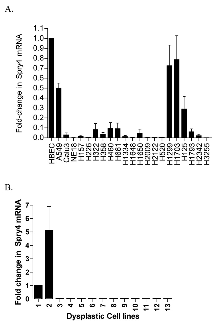
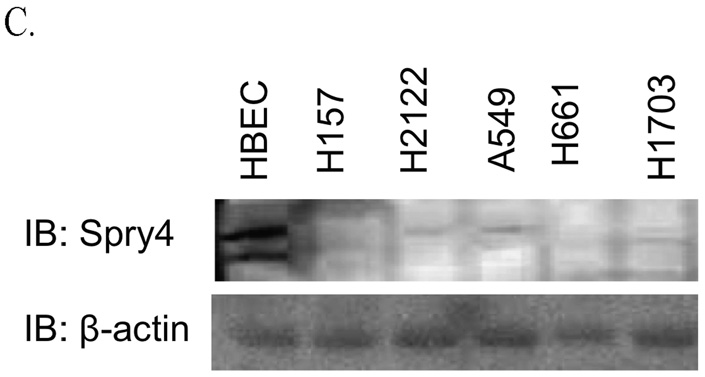
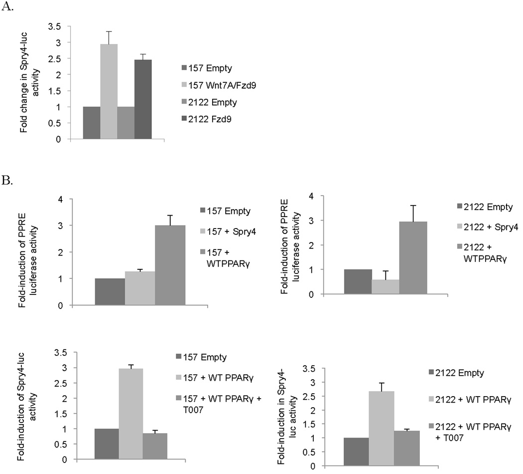
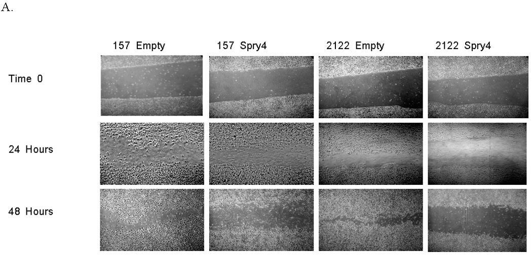
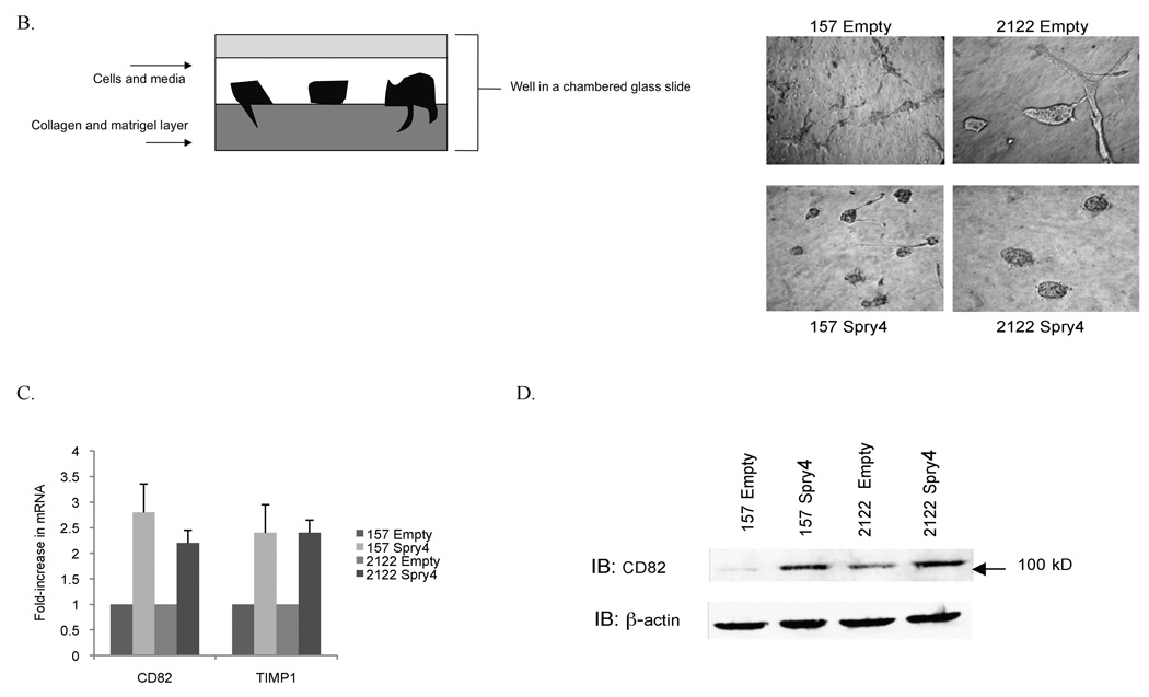
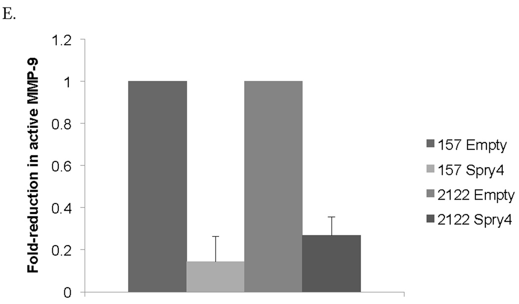
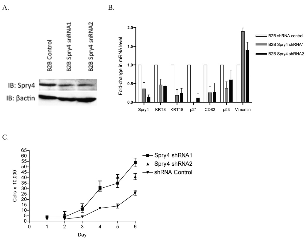
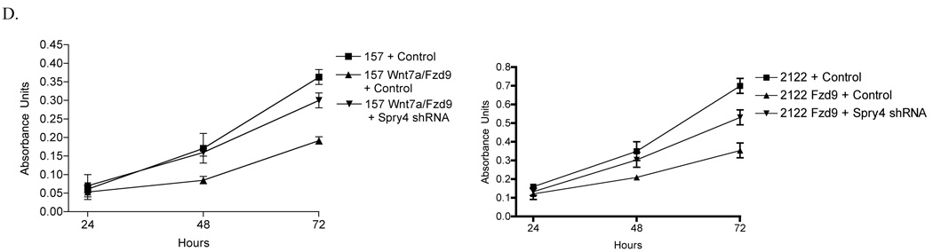
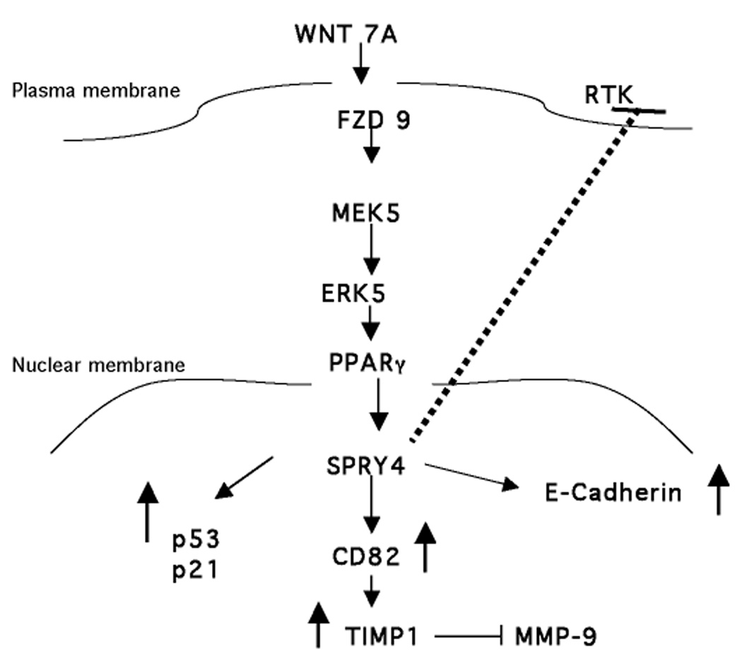
Sprouty proteins are potent receptor tyrosine kinase inhibitors that antagonize growth factor signaling and are involved in lung development. However, little is known about the regulation or targets of Sprouty-4 (Spry4) in lung cancer. Our study aimed to determine the role of Spry4 in non-small cell lung cancer (NSCLC). We found that Spry4 mRNA expression was decreased in NSCLC cell lines and in dysplastic lung cell lines compared with a nontransformed cell line, suggesting that Spry4 has tumor-suppressing activity. When Spry4 was stably transfected into H157 and H2122 NSCLC cell lines, decreased migration and invasion were observed. Matrix metalloproteinase-9 activity was decreased, and the expression of matrix metalloproteinase inhibitors TIMP1 and CD82 were increased. Stable expression of Spry4 led to reduced cell growth and reduced anchorage-independent growth in NSCLC cell lines, along with upregulation of tumor suppressors p53 and p21. Changes in epithelial and mesenchymal markers indicated that Spry4 expression induces a reversal of the epithelial to mesenchymal transition characteristic of tumor cells. Treatment of a nontransformed lung epithelial cell line with short hairpin RNA to Spry4 led to the decreased expression of epithelial markers and increased cell growth, supporting the concept of Spry4 acting as a tumor suppressor. We showed that the activity of the Spry4 promoter is increased by Wnt7A/Fzd9 signaling through peroxisome proliferator-activated receptor gamma. These data present previously undescribed targets of Spry4 and suggest that Spry4 is a downstream target of Wnt7A/Fzd 9 signaling. Spry4 may have efficacy in the treatment of NSCLC.


Figures

Figure 1
Figure 1
Spry4 expression is decreased in NSCLC cell lines and dysplastic lung cell culture. (A,B) QPCR for Spry4 mRNA in NSCLC cell lines and dysplastic lung epithelial cell culture. Dysplastic cell cultures are arbitrarily numbered 1–13. Cell line mRNA levels are presented as fold-reductions compared to the non-transformed lung epithelial cell line HBEC. Results are the average of triplicate experiments normalized to GAPDH. (C) Immunoblot analysis of Spry4 protein expression in NSCLC cell lines compared to a non-transformed lung epithelial cell line (HBEC). β-actin is included as a loading control.
Figure 1
Figure 1
Spry4 expression is decreased in NSCLC cell lines and dysplastic lung cell culture. (A,B) QPCR for Spry4 mRNA in NSCLC cell lines and dysplastic lung epithelial cell culture. Dysplastic cell cultures are arbitrarily numbered 1–13. Cell line mRNA levels are presented as fold-reductions compared to the non-transformed lung epithelial cell line HBEC. Results are the average of triplicate experiments normalized to GAPDH. (C) Immunoblot analysis of Spry4 protein expression in NSCLC cell lines compared to a non-transformed lung epithelial cell line (HBEC). β-actin is included as a loading control.
Figure 2
Figure 2
The Spry4 promoter is activated by PPARγ expression. (A) H157 and H2122 cells with stable Wnt7A and/or Fzd9 expression were transfected with Spry4-luciferase. Results are the average of triplicate experiments. (B) H157 and H2122 cells were transiently transfected with PPAR response element (PPRE) –luc, Spry4 and wild type PPARγ. H157 and H2122 cells were transfected with wild type PPARγ and Spry4 promoter luciferase and treated with T007, a PPARγ inhibitor. Results are the average of triplicate experiments. (C) H157 and H2122 cells with stable PPARγ expression were transfected with full length (−4446) and truncated versions of Spry4-luciferase (−1182, −418, −31). Results are the average of triplicate experiments. Change in activity is presented as fold-induction of the luciferase reporter.
Figure 2
Figure 2
The Spry4 promoter is activated by PPARγ expression. (A) H157 and H2122 cells with stable Wnt7A and/or Fzd9 expression were transfected with Spry4-luciferase. Results are the average of triplicate experiments. (B) H157 and H2122 cells were transiently transfected with PPAR response element (PPRE) –luc, Spry4 and wild type PPARγ. H157 and H2122 cells were transfected with wild type PPARγ and Spry4 promoter luciferase and treated with T007, a PPARγ inhibitor. Results are the average of triplicate experiments. (C) H157 and H2122 cells with stable PPARγ expression were transfected with full length (−4446) and truncated versions of Spry4-luciferase (−1182, −418, −31). Results are the average of triplicate experiments. Change in activity is presented as fold-induction of the luciferase reporter.
Figure 3
Figure 3
Stable expression of Spry4 reduces cell growth and anchorage independent growth in NSCLC cells. (A) Stable Spry4 expression in transfected H157 and H2122 cells was verified by immunoblot with a GAPDH loading control and by QPCR. (B) 50,000 cells from H157 and H2122 stably expressing Spry4 or an empty vector control were plated in triplicate in 6 wells and one well was counted each day. Results are the average of triplicate experiments. (C) 25,000 cells from H157 and H2122 stably expressing Spry4 or an empty vector control were seeded in triplicate in media with 0.3% agar on a base of 0.5% agar. Colonies were stained with nitroblue tetrazolium chloride at 21 days. Representative soft agar pictures are shown. Cloning efficiency is the ratio of counted colonies to seeded cells and is the average of triplicate experiments. (D) QPCR for p21 in H2122 cell lines stably expressing Spry4 compared to an empty vector control. Results are the average of triplicate experiments normalized to GAPDH.
Figure 3
Figure 3
Stable expression of Spry4 reduces cell growth and anchorage independent growth in NSCLC cells. (A) Stable Spry4 expression in transfected H157 and H2122 cells was verified by immunoblot with a GAPDH loading control and by QPCR. (B) 50,000 cells from H157 and H2122 stably expressing Spry4 or an empty vector control were plated in triplicate in 6 wells and one well was counted each day. Results are the average of triplicate experiments. (C) 25,000 cells from H157 and H2122 stably expressing Spry4 or an empty vector control were seeded in triplicate in media with 0.3% agar on a base of 0.5% agar. Colonies were stained with nitroblue tetrazolium chloride at 21 days. Representative soft agar pictures are shown. Cloning efficiency is the ratio of counted colonies to seeded cells and is the average of triplicate experiments. (D) QPCR for p21 in H2122 cell lines stably expressing Spry4 compared to an empty vector control. Results are the average of triplicate experiments normalized to GAPDH.
Figure 4
Figure 4
Spry4 re-expression induces a more epithelial phenotype in NSCLC cells. (A) 5,000 cells from H157 and H2122 stably expressing Spry4 or an empty vector control were seeded in media with 4% matrigel on top of a 1:1 matrigel and media base layer. Cell morphology was recorded on day 5. Data is representative of triplicate experiments. (B) Western blot analysis of E-cadherin in cell lysates from H157 and H2122 cell lines with stable Spry4 expression compared to an empty vector control. Loading control is β-actin and data are representative of triplicate experiments. (C) QPCR for KRT8, KRT18, and Vimentin in H157 and H2122 cell lines stably expressing Spry4 compared to an empty vector control. Results are the average of triplicate experiments and are normalized to GAPDH.
Figure 5
Figure 5
Re-expression of Spry4 inhibits migration and invasion in NSCLC cells. (A) A 3mm space was created across the diameter of plates of H157 and H2122 cells stably expressing Spry4 or an empty vector control and migration was recorded at 24 and 48 hours. Results are representative of triplicate experiments. (B) The 3D invasion assay uses a layer of collagen beneath a layer of matrigel and media containing the cells of interest. 5,000 cells from H157 and H2122 stably expressing Spry4 or an empty vector control were seeded in triplicate in media with 4% matrigel on top of a 1:1 matrigel and collagen base layer. Cell invasion was recorded on day 5. (C) Expression levels of CD82 and TIMP1 were assessed by QPCR in H157 and H2122 cell lines stably expressing Spry4 or an empty vector control. Results are the average of triplicate experiments normalized to GAPDH. (D) Increased expression of CD82 was confirmed by western blot analysis of cell lysates from H157 and H2122 cell lines stably expressing Spry4 compared to an empty vector control. Loading control is β-actin. (e) H157 cells stably expressing Spry4, H2122 cells stably expressing Spry4, and corresponding empty vector controls were seeded in a 96-well plate and analyzed with the MMP-9 Human Biotrak Assay. Absorbance was measured and a standard curve was used to calculate the amount of active MMP-9 in the samples. Results are the average of triplicate experiments with standard error.
Figure 5
Figure 5
Re-expression of Spry4 inhibits migration and invasion in NSCLC cells. (A) A 3mm space was created across the diameter of plates of H157 and H2122 cells stably expressing Spry4 or an empty vector control and migration was recorded at 24 and 48 hours. Results are representative of triplicate experiments. (B) The 3D invasion assay uses a layer of collagen beneath a layer of matrigel and media containing the cells of interest. 5,000 cells from H157 and H2122 stably expressing Spry4 or an empty vector control were seeded in triplicate in media with 4% matrigel on top of a 1:1 matrigel and collagen base layer. Cell invasion was recorded on day 5. (C) Expression levels of CD82 and TIMP1 were assessed by QPCR in H157 and H2122 cell lines stably expressing Spry4 or an empty vector control. Results are the average of triplicate experiments normalized to GAPDH. (D) Increased expression of CD82 was confirmed by western blot analysis of cell lysates from H157 and H2122 cell lines stably expressing Spry4 compared to an empty vector control. Loading control is β-actin. (e) H157 cells stably expressing Spry4, H2122 cells stably expressing Spry4, and corresponding empty vector controls were seeded in a 96-well plate and analyzed with the MMP-9 Human Biotrak Assay. Absorbance was measured and a standard curve was used to calculate the amount of active MMP-9 in the samples. Results are the average of triplicate experiments with standard error.
Figure 6
Figure 6
Treatment of a non-transformed lung epithelial cell line, Beas2B (B2B), with shRNA to Spry4 leads to changes in EMT markers and increased cell growth. (A) Immunoblot for expression of Spry4 and E-cadherin in B2B cells treated with shRNA to Spry4. Loading control is βactin. (B) QPCR was used to measure Spry4, KRT8, KRT18, CD82, p53, p21, and Vimentin mRNA levels in B2B cells treated with Spry4 shRNA. Results are the average of triplicate experiments normalized to GAPDH. (C) 20,000 cells from B2B cells treated with shRNA to Spry4 or an shRNA control were plated in 6 wells and one well was counted each day. Data shown is the average of triplicate experiments. (D) 500 cells from each cell line were seeded per well in triplicate. At 24, 48, and 72 hours, 20ul of MTS reagent (Promega) was added to each well, incubated for 1 hour at 37C, and analyzed at 490nm. Results are presented as the average of triplicate experiments.
Figure 6
Figure 6
Treatment of a non-transformed lung epithelial cell line, Beas2B (B2B), with shRNA to Spry4 leads to changes in EMT markers and increased cell growth. (A) Immunoblot for expression of Spry4 and E-cadherin in B2B cells treated with shRNA to Spry4. Loading control is βactin. (B) QPCR was used to measure Spry4, KRT8, KRT18, CD82, p53, p21, and Vimentin mRNA levels in B2B cells treated with Spry4 shRNA. Results are the average of triplicate experiments normalized to GAPDH. (C) 20,000 cells from B2B cells treated with shRNA to Spry4 or an shRNA control were plated in 6 wells and one well was counted each day. Data shown is the average of triplicate experiments. (D) 500 cells from each cell line were seeded per well in triplicate. At 24, 48, and 72 hours, 20ul of MTS reagent (Promega) was added to each well, incubated for 1 hour at 37C, and analyzed at 490nm. Results are presented as the average of triplicate experiments.
Figure 7
Figure 7
A model for Wnt7A/Fzd9 activation of Spry4 through PPARγ. Expression of Spry4 results in decreased MMP-9 activity, through CD82 and TIMP1, and increased E-cadherin, p21, and p53 expression.

Similar articles

Cited by

References

    1. Hacohen N, Kramer S, Sutherland D, Hiromi Y, Krasnow MA. sprouty encodes a novel antagonist of FGF signaling that patterns apical branching of the Drosophila airways. Cell. 1998;92(2):253–263. - PubMed
    1. Casci T, Vinos J, Freeman M. Sprouty, an intracellular inhibitor of Ras signaling. Cell. 1999;96(5):655–665. - PubMed
    1. Mason JM, Morrison DJ, Basson MA, Licht JD. Sprouty proteins: multifaceted negative-feedback regulators of receptor tyrosine kinase signaling. Trends Cell Biol. 2006;16(1):45–54. - PubMed
    1. Gross I, Bassit B, Benezra M, Licht JD. Mammalian sprouty proteins inhibit cell growth and differentiation by preventing ras activation. J Biol Chem. 2001;276(49):46460–46468. - PubMed
    1. Sasaki A, Taketomi T, Kato R, et al. Mammalian Sprouty4 suppresses Ras-independent ERK activation by binding to Raf1. Nat Cell Biol. 2003;5(5):427–432. - PubMed

Publication types

MeSH terms