. 2010 Aug;151(8):3566-76.
doi: 10.1210/en.2010-0204. Epub 2010 May 25.

Impaired-inactivation of FoxO1 contributes to glucose-mediated increases in serum very low-density lipoprotein

Affiliations

Impaired-inactivation of FoxO1 contributes to glucose-mediated increases in serum very low-density lipoprotein

Ke Wu et al. Endocrinology. 2010 Aug.

Abstract

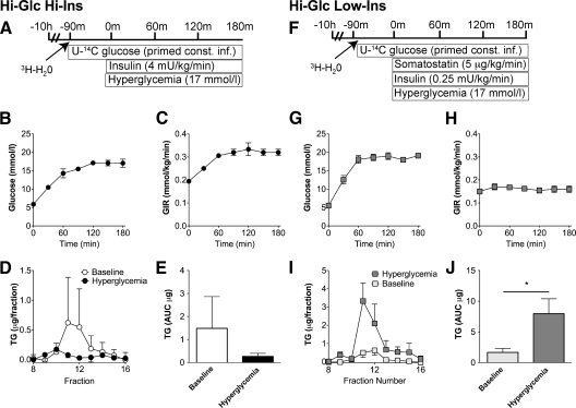
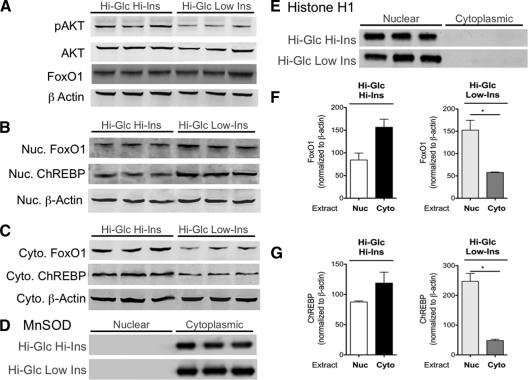
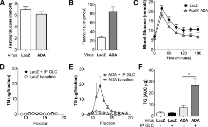
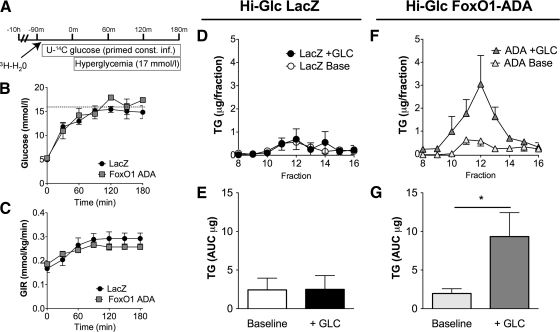
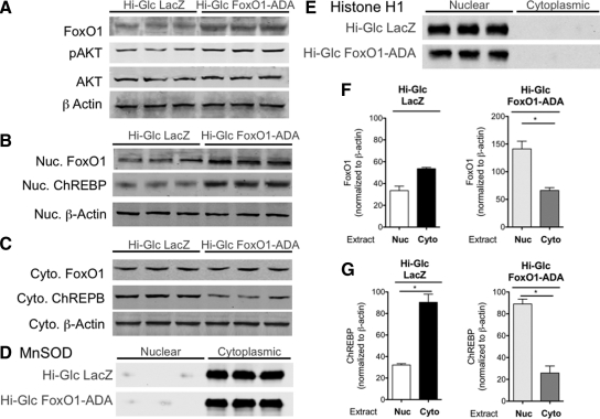
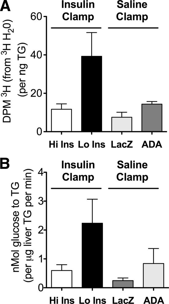
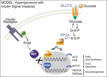
For patients with diabetes, insulin resistance and hyperglycemia both contribute to increased serum triglyceride in the form of very low-density lipoprotein (VLDL). Our objective was to define the insulin conditions in which hyperglycemia promotes increased serum VLDL in vivo. We performed hyperglycemic-hyperinsulinemic clamp studies and hyperglycemic-hypoinsulinemic clamp studies in rats, with metabolic tracers for glucose flux and de novo fatty acid synthesis. When blood glucose was clamped at hyperglycemia (17 mm) for 2 h under hyperinsulinemic conditions (4 mU/kg . min), serum VLDL levels were not increased compared with baseline. We speculated that hyperinsulinemia minimized glucose-mediated VLDL changes and performed hyperglycemic-hypoinsulinemic clamp studies in which insulin was clamped near fasting levels with somatostatin (17 mm blood glucose, 0.25 mU/kg . min insulin). Under low-insulin conditions, serum VLDL levels were increased 4.7-fold after hyperglycemia, and forkhead box O1 (FoxO1) was not excluded from the nucleus of liver cells. We tested the extent that impaired inactivation of FoxO1 by insulin was sufficient for glucose to promote increased serum VLDL. We found that, when the ability of insulin to inactivate FoxO1 is blocked after adenoviral delivery of constitutively active FoxO1, glucose increased serum VLDL triglyceride when given both by ip glucose tolerance testing (3.5-fold increase) and by a hyperglycemic clamp (4.6-fold). Under both experimental conditions in which insulin signaling to FoxO1 was impaired, we found increased activation of carbohydrate response element binding protein. These data suggest that glucose more potently promotes increased serum VLDL when insulin action is impaired, with either low insulin levels or disrupted downstream signaling to the transcription factor FoxO1.


Figures

Figure 1
Figure 1
A–E, Hyperglycemic-hyperinsulinemic clamp. A, Study design. B, Blood glucose during clamp studies. C, GIR during clamp. D, TG content of VLDL fractions after HPLC. E, AUC VLDL-TG. F–J, Hyperglycemic-hypoinsulinemic clamp. F, Study design. G, Blood glucose during clamp studies. H, GIR during clamp. I, TG content of VLDL fractions after HPLC. J, AUC VLDL-TG. *, P < 0.05. Glc, Glucose; Ins, insulin.
Figure 2
Figure 2
Insulin signaling, high-insulin, and low-insulin clamp studies. A, Western blots for AKT, phosphorylated AKT (pAKT), and FoxO1 from whole-cell extracts of liver after clamp studies. B, Western blots for FoxO1 and ChREBP from nuclear extracts from liver. C, Western blots for FoxO1 and ChREBP from cytoplasmic extracts from liver. D, Western blots of nuclear and cytoplasmic extracts for MnSOD, a cytoplasmic marker. E, Western blots of nuclear and cytoplasmic extracts for histone H1, a nuclear marker. F, Quantitation of FoxO1 in nuclear and cytoplasmic compartments normalized to β-actin in those fractions. G, Quantitation of ChREBP in nuclear and cytoplasmic compartments normalized to β-actin in those fractions. *, P < 0.05. Nuc., Nuclear; Cyto., cytoplasmic; Glc, glucose; Ins, insulin.
Figure 3
Figure 3
Intraperitoneal GTT after adenovirus-mediated gene expression. A, Ten-hour fasting glucose level 5 d after 1 × 10⁹ pfu injection of adenovirus-expressing LacZ (LacZ) or adenovirus-expressing FoxO1-ADA (ADA). B, Fasting insulin. C, Blood glucose after ip GTT with glucose given at 3 g/kg lean mass. D, Serum VLDL-TG after adenovirus LacZ expression. E, Serum VLDL-TG after adenovirus FoxO1-ADA expression. F, AUC VLDL from D and E. *, P < 0.05. GLC, Glucose.
Figure 4
Figure 4
Hyperglycemic clamp after adenovirus-mediated gene expression: A, Study design for clamp performed after a 10-h fast, 5 d after 1 × 10⁹ pfu injection of adenovirus-expressing LacZ or adenovirus-expressing FoxO1-ADA. B, Blood glucose during clamp. C, GIR. D, TG content of VLDL fractions for the LacZ group. E, VLDL AUC for the LacZ group. F, TG content of VLDL fractions for the ADA group. G, VLDL AUC for the ADA group. *, P < 0.05. GLC, Glucose.
Figure 5
Figure 5
Insulin signaling, LacZ-FoxO1-ADA studies. A, Western blots for AKT, phosphorylated AKT (pAKT), and FoxO1 from whole-cell extracts of liver after clamp studies. B, Western blots for FoxO1 and ChREBP from nuclear extracts from liver. C, Western blots for FoxO1 and ChREBP from cytoplasmic extracts from liver. D, Western blots of nuclear and cytoplasmic extracts for MnSOD, a cytoplasmic marker. E, Western blots of nuclear and cytoplasmic extracts for histone H1, a nuclear marker. F, Quantitation of FoxO1 in nuclear and cytoplasmic compartments normalized to β-actin in those fractions. G, Quantitation of ChREBP in nuclear and cytoplasmic compartments normalized to β-actin in those fractions. *, P < 0.05. Nuc., Nuclear; Cyto., cytoplasmic; Glc, glucose; Ins, insulin.
Figure 6
Figure 6
Tracer flux to hepatic lipid. A, 3H incorporation into hepatic TG (from [3H]H2O). B, Rate of glucose incorporation into hepatic TG.
Figure 7
Figure 7
Real-time PCR of liver mRNA normalized to cyclophilin expression. A–E, High-insulin and low-insulin clamp studies. A, B, Gluconeogenesis, PEPCK and G6Pase. C–E, Fatty acid synthesis, ACC and FAS; VLDL assembly, Mttp. F–J, LacZ and FoxO1-ADA studies. F, G, Gluconeogenesis, PEPCK and G6Pase. H–J, Fatty acid synthesis. ACC and FAS; VLDL assembly, Mttp. Error bars represent sd. *, P < 0.05. Ins, Insulin.
Figure 8
Figure 8
Model. When insulin signaling is unable to phosphorylate FoxO1, FoxO1 remains in the nuclear compartment to increase the expression of genes whose products promote gluconeogenesis and VLDL assembly. Impaired insulin signaling to FoxO1 may also result in increased glucose-mediated activation of ChREBP, which promotes expression of genes whose products promote fatty acid synthesis. GLUT2, Glucose transporter 2; G-6-P, glucose-6-phosphate; PP2A, protein phosphatase 2A; IRS, insulin receptor substrate; PI3K, phosphatidylinositol 3-kinase; x-5-p, xylulose-5-phosphate.

Similar articles

Cited by

References

    1. Adiels M, Borén J, Caslake MJ, Stewart P, Soro A, Westerbacka J, Wennberg B, Olofsson SO, Packard C, Taskinen MR 2005 Overproduction of VLDL1 driven by hyperglycemia is a dominant feature of diabetic dyslipidemia. Arterioscler Thromb Vasc Biol 25:1697–1703 - PubMed
    1. Bernstein RM, Davis BM, Olefsky JM, Reaven GM 1978 Hepatic insulin responsiveness in patients with endogenous hypertriglyceridaemia. Diabetologia 14:249–253 - PubMed
    1. Gill JM, Brown JC, Bedford D, Wright DM, Cooney J, Hughes DA, Packard CJ, Caslake MJ 2004 Hepatic production of VLDL1 but not VLDL2 is related to insulin resistance in normoglycaemic middle-aged subjects. Atherosclerosis 176:49–56 - PubMed
    1. Kissebah AH, Alfarsi S, Adams PW, Wynn V 1976 Role of insulin resistance in adipose tissue and liver in the pathogenesis of endogenous hypertriglyceridaemia in man. Diabetologia 12:563–571 - PubMed
    1. Haffner SM, Valdez RA, Hazuda HP, Mitchell BD, Morales PA, Stern MP 1992 Prospective analysis of the insulin-resistance syndrome (syndrome X). Diabetes 41:715–722 - PubMed

Publication types

MeSH terms