. 2010 Jun;9(6):1638-46.
doi: 10.1158/1535-7163.MCT-10-0097. Epub 2010 May 25.

Global targeting of subcellular heat shock protein-90 networks for therapy of glioblastoma

Affiliations

Global targeting of subcellular heat shock protein-90 networks for therapy of glioblastoma

Markus D Siegelin et al. Mol Cancer Ther. 2010 Jun.

Abstract

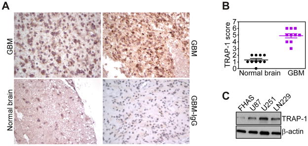
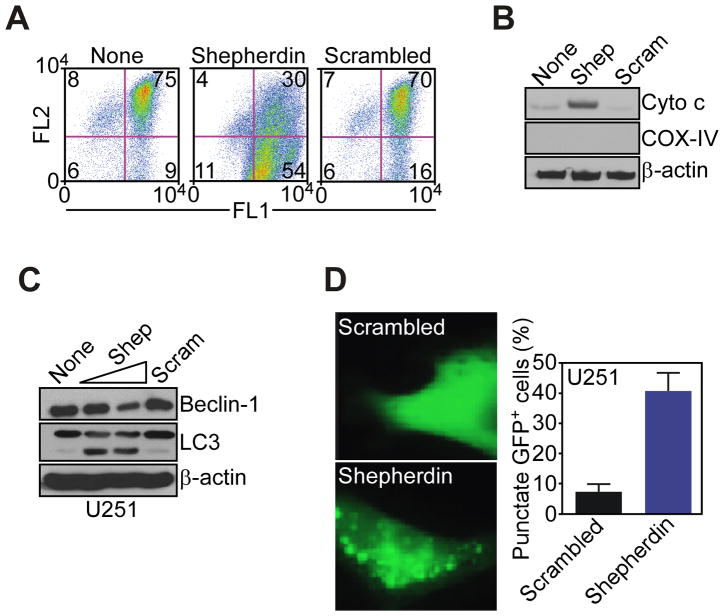
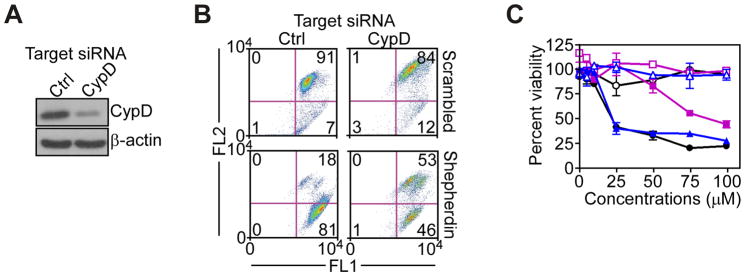
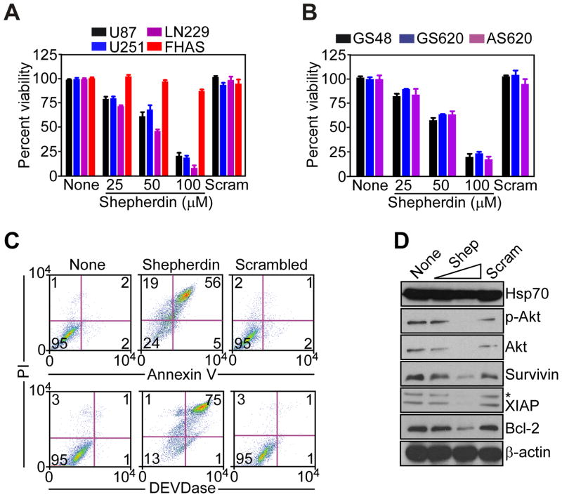
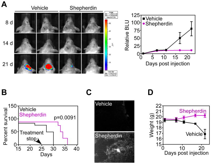
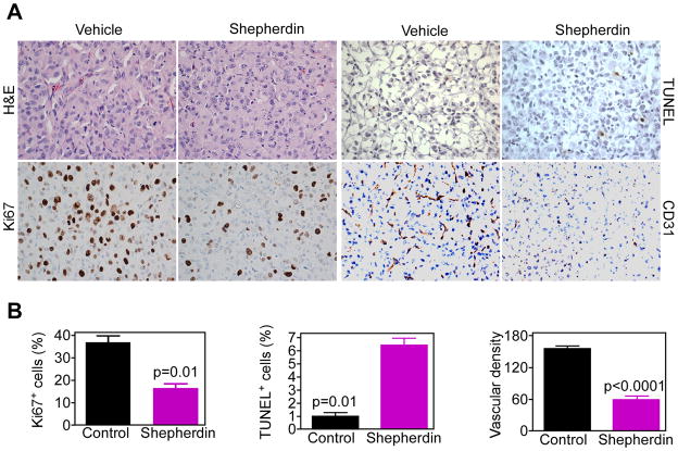
Drug discovery for complex and heterogeneous tumors now aims at dismantling global networks of disease maintenance, but the subcellular requirements of this approach are not understood. Here, we simultaneously targeted the multiple subcellular compartments of the molecular chaperone heat shock protein-90 (Hsp90) in a model of glioblastoma, a highly lethal human malignancy in urgent need of fresh therapeutic strategies. Treatment of cultured or patient-derived glioblastoma cells with Shepherdin, a dual peptidomimetic inhibitor of mitochondrial and cytosolic Hsp90, caused irreversible collapse of mitochondria, degradation of Hsp90 client proteins in the cytosol, and tumor cell killing by apoptosis and autophagy. Stereotactic or systemic delivery of Shepherdin was well tolerated and suppressed intracranial glioma growth via inhibition of cell proliferation, induction of apoptosis, and reduction of angiogenesis in vivo. These data show that disabling Hsp90 cancer networks in their multiple subcellular compartments improves strategies for drug discovery and may provide novel molecular therapy for highly recalcitrant human tumors.


Conflict of interest statement

Conflict of Interest: The authors declare that no conflict of interest exists.

Figures

Figure 1
Figure 1
Differential expression of TRAP-1 in human gliomas. A, representative cases of human glioblastoma (GBM), or adjacent normal brain were stained with an antibody to TRAP-1 or IgG (GBM-IgG), and analyzed by immunohistochemistry. Magnification, x100, x200. B, quantification of TRAP-1 expression. The indicated samples of glioblastoma (GBM) or adjacent normal brain were scored for TRAP-1 staining intensity and percentage of positive cells. C, extracts from U87, U251 or LN229 glioblastoma cells or normal fetal human astrocytes (FHAS) were analyzed by Western blotting.
Figure 2
Figure 2
Shepherdin-induced mitochondrial dysfunction and autophagy. A, glioblastoma LN229 cells were incubated with Shepherdin or scrambled peptidomimetic and analyzed for mitochondrial membrane potential by multi-parametric flow-cytometry after 3 h. The percentage of cells in each quadrant is indicated. FL1, green fluorescence channel; FL2: red fluorescence channel. B, LN229 cells incubated with Shepherdin (Shep) or scrambled peptidomimetic (Scram) were analyzed by Western blotting after 3 h. C, glioblastoma U251 cells treated with Shepherdin (Shep, 50–100 μM) or scrambled peptidomimetic (Scram, 100 μM) were analyzed by Western blotting. D, U251 cells transfected with LC3-GFP cDNA were treated with Shepherdin or scrambled peptidomimetic, and analyzed by fluorescence microscopy. Right, quantification of cells with punctate GFP staining.
Figure 3
Figure 3
Requirement of CypD in Shepherdin-induced mitochondrial dysfunction. A, LN229 cells were transfected with control, non-targeting or CypD-directed siRNA, and analyzed by Western blotting. B, LN229 cells transfected with the indicated siRNA were analyzed for changes in mitochondrial membrane potential after 3 h. The percentage of cells in each quadrant is indicated. FL1, green fluorescence channel; FL2, red fluorescence channel. C, LN229 cells transfected with non-targeting (blue) or CypD (purple)-directed siRNA were left untreated (black) or incubated with Shepherdin (solid symbols) or scrambled peptidomimetic (open symbols), and analyzed by MTT. Data are the mean±SEM.
Figure 4
Figure 4
Multimodal antiglioma activity of Shepherdin. A, B, The indicated glioblastoma cell lines or FHAS (A), or patient-derived, primary glioblastoma cultures (B), were left untreated (None) or incubated with increasing concentrations of Shepherdin or scrambled peptidomimetic (Scram), and analyzed by MTT after 3 h. Data are the mean±SEM, n=3. C, untreated (None) or Shepherdin- or scrambled peptidomimetic-treated U87 cells were analyzed after 16 h for Annexin V/PI staining (top), or DEVDase (caspase)/PI activity (bottom), by multiparametric flow cytometry. The percentage of cells in each quadrant is indicated. D, U87 cells treated with Shepherdin (Shep, 50–100 μM) or scrambled peptidomimetic (Scram, 100 μM) were analyzed by Western blotting. p-Akt, Ser473-phosphorylated Akt. *, non-specific.
Figure 5
Figure 5
Anti-glioma activity of Shepherdin, in vivo. A, SCID/beige mice stereotactically implanted with U87-Luc cells in the right cerebral striatum were treated systemically with vehicle or Shepherdin (50 mg/kg daily i.p.), and imaged by live bioluminescence at the indicated time intervals. Representative animals per group are shown. Right, quantification of bioluminescence units (BLU) in vehicle or Shepherdin-treated groups. Data are the mean±SD of the various groups (5 animals/group). B, survival of SCID/beige mice implanted with U87-Luc cells and treated systemically with vehicle or Shepherdin (50 mg/kg daily i.p.). C, SCID/beige mice implanted with U87-Luc cells were injected with biotin-conjugated Shepherdin or vehicle, and brain samples were analyzed for intracranial accumulation of Shepherdin by fluorescence microscopy after 24 h. D, weight changes in vehicle- or Shepherdin-treated mice. Data are the mean±SD of the various groups (5 animals/group).
Figure 6
Figure 6
Histopathology of Shepherdin anti-glioma activity, in vivo. A, representative brain sections from vehicle- or Shepherdin-treated mice were harvested after 20 d, and analyzed by H&E, cell proliferation (Ki67), apoptosis (TUNEL), or angiogenesis (CD31), by immunohistochemistry. Magnification, x400. B, quantification of mitotic index (left), apoptosis (middle) and microvessel density (right). Labeled cells were counted in an average of 4–6 high- power fields. Data are mean±SEM.

References

    1. Vogelstein B, Kinzler KW. Cancer genes and the pathways they control. Nat Med. 2004;10:789–99. - PubMed
    1. Wood LD, Parsons DW, Jones S, et al. The genomic landscapes of human breast and colorectal cancers. Science. 2007;318:1108–13. - PubMed
    1. Luo J, Solimini NL, Elledge SJ. Principles of cancer therapy: oncogene and non-oncogene addiction. Cell. 2009;136:823–37. - PMC - PubMed
    1. Stein WD, Bates SE, Fojo T. Intractable cancers: the many faces of multidrug resistance and the many targets it presents for therapeutic attack. Curr Drug Targets. 2004;5:333–46. - PubMed
    1. Purow B, Schiff D. Advances in the genetics of glioblastoma: are we reaching critical mass? Nat Rev Neurol. 2009;5:419–26. - PMC - PubMed

Publication types

MeSH terms