. 2010 Jun 15;70(12):5034-45.
doi: 10.1158/0008-5472.CAN-10-0563. Epub 2010 May 25.

Mechanism by which Mcl-1 regulates cancer-specific apoptosis triggered by mda-7/IL-24, an IL-10-related cytokine

Affiliations

Mechanism by which Mcl-1 regulates cancer-specific apoptosis triggered by mda-7/IL-24, an IL-10-related cytokine

Rupesh Dash et al. Cancer Res. .

Abstract

Melanoma differentiation-associated gene-7/interleukin-24 (mda-7/IL-24), a cytokine belonging to the IL-10 family, selectively induces apoptosis in cancer cells without harming normal cells by promoting an endoplasmic reticulum (ER) stress response. The precise molecular mechanism by which the ER stress response culminates in cell death requires further clarification. The present study shows that in prostate carcinoma cells, the mda-7/IL-24-induced ER stress response causes apoptosis by translational inhibition of the antiapoptotic protein myeloid cell leukemia-1 (Mcl-1). Forced expression of Mcl-1 blocked mda-7/IL-24 lethality, whereas RNA interference or gene knockout of Mcl-1 markedly sensitized transformed cells to mda-7/IL-24. Mcl-1 downregulation by mda-7/IL-24 relieved its association with the proapoptotic protein Bak, causing oligomerization of Bak and leading to cell death. These observations show the profound role of the Bcl-2 protein family member Mcl-1 in regulating cancer-specific apoptosis induced by this cytokine. Thus, our studies provide further insights into the molecular mechanism of ER stress-induced cancer-selective apoptosis by mda-7/IL-24. As Mcl-1 is overexpressed in the majority of prostate cancers, mda-7/IL-24 might provide an effective therapeutic for this disease.


Figures

Figure 1
Figure 1
Mcl-1 is overexpressed in prostate carcinomas. A, cell lysates were collected from the indicated cells, and expression of Mcl-1 was determined by Western blotting. Immunohistologic analysis of Mcl-1 expression in tissue microarrays: normal human prostate tissue (B); stage I (C), stage II (D), stage III (E), and stage IV (F) prostate cancer tissues (magnification, ×200).
Figure 2
Figure 2
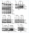
Forced expression of Mcl-1 blocks mda-7/IL-24-mediated apoptosis in DU-145 cells in vitro and in vivo. A, DU-145 cells were infected with 100 pfu/cell of Ad.vec or Ad.mda-7, protein lysates were prepared at the specified time points, and expression profiles of the indicated proteins were determined by Western blotting. B, DU-145 cells were infected with the indicated pfu/cell of Ad.vec or Ad.mda-7 for 48 h, and expression of Mcl-1 and MDA-7/IL-24 and activation of eIF2α were determined by Western blotting. C, DU-145 cells were transfected with a dominant-negative PERK (PERK DN) or control vector and then infected with the indicated pfu/cell of either Ad.vec or Ad.mda-7 for 48 h. Expression of the indicated proteins was determined by Western blotting. D, DU-145 cells were transfected with an eIF2αDN or control vector and then infected with indicated pfu/cell of either Ad.vec or Ad.mda-7 for 48 h. Expression of the indicated proteins was determined by Western blotting. E, PC-3 prostate cancer cells were infected with the indicated pfu/cell of Ad.5-vec, Ad.5/3-vec, Ad.5-mda-7, or Ad.5/3-mda-7, and expression of Mcl-1 and MDA-7/IL-24 was determined by Western blotting. F, normal prostate epithelial cells (P69) were infected with the indicated pfu/cell of Ad.vec or Ad.mda-7, and expression of Mcl-1 and MDA-7/IL-24 was determined by Western blotting. Percentage of apoptosis in A to F was determined by Annexin V–fluorescein isothiocyanate/propidium iodide (Annexin V–FITC/PI) staining. Bars, SD (n =3).
Figure 3
Figure 3
Forced expression of Mcl-1 blocks mda-7/IL-24-mediated apoptosis in DU-145 cells in vitro and in vivo. A, DU-145 cells were transiently transfected with pcDNA3.1 or pcDNA3.1-Mcl-1 plasmids, cultured for 20 h, then infected with the indicated pfu/cell of Ad.vec or Ad.mda-7 for 48 h, after which protein lysates were prepared and subjected to Western blotting using the indicated antibody. B, DU-145 cells stably expressing (inset) a transfected Mcl-1 gene (two clones, DU-Mcl-1-8 (1,6-bismaleimidohexane) and DU-Mcl-1-20) or an empty vector (DU-pcDNA3.1) were infected with the indicated pfu/cell of Ad.vec or Ad.mda-7 for 48 h, after which the extent of apoptosis was determined using Annexin V staining assays. Bars, SD (n =3;*, P < 0.05 versus DU-pcDNA3.1). C, lysates were prepared from DU-Mcl-1-8 or DU-pcDNA3.1 cells infected with the indicated pfu/cell of Ad.vec or Ad.mda-7 for 48 h, and Western blotting was performed to monitor Mcl-1 and MDA-7/IL-24 protein levels and cleavage of PARP. D, tumor xenografts from DU-Mcl-1-8 and DU-pcDNA3.1 cells were established in the left flanks of athymic nude mice, and the tumors were injected with the indicated adenoviruses (100 pfu/cell) over a 4-wk period (total of seven injections) and tumor size was measured. Bars, SD (n = 5 mice). E, measurement of tumor weight at the end of the study. Bars, SD (n = 5 mice). Photograph of the DU-Mcl-1-8 and DU-pcDNA3.1 xenograft tumors after 5 wk (inset). F, tumor xenograft models established as indicated in B. Tumors were harvested, paraffin-embedded sections were immunostained for MDA-7/IL-24, and Ki-67 and TUNEL assays were performed using Deadend TUNEL assay kit.
Figure 4
Figure 4
Forced expression of mda-7/IL-24 promotes Mcl-1 degradation to trigger Bak activation. A, lysates were prepared from DU-Mcl-1-8, a clone of DU-145 cells ectopically expressing Mcl-1, or DU-pcDNA3.1, an empty vector, and infected with the indicated pfu/cell of Ad.vec or Ad.mda-7 for 48 h; expression of Bak was determined by Western blotting. For detection of activated Bak, cell lysates were immunoprecipitated with Bak and Western blotting was performed with anti-Bak Ab-1 that specifically recognizes activated Bak. B, cells were treated as mentioned in A, and Bak activation was detected by flow cytometric analysis of Ad.vec- or Ad.mda-7 (100 pfu/cell for 48 h)–infected DU-Mcl-1-8 and DU-pcDNA3.1 cells stained with anti-Bak Ab-1 antibody. C, DU-Mcl-1-8 and DU-pcDNA3.1 cells were infected with the indicated pfu/cell of Ad.vec or Ad.mda-7 for 48 h, and the mitochondrial fractions were isolated, treated with 1 mmol/L BMH, and then analyzed by SDS-PAGE and immunoblotted with anti-BAK. *, BAK complexes with dimers or trimers. **, inactive BAK conformer. D, DU-Mcl-1-8 (1,6-bismaleimidohexane) and DU-pcDNA3.1 cells were infected with the indicated pfu/cell of Ad.vec and Ad.mda-7 for 24 h in the absence or presence of 25 μmol/L of z-VAD-FMK, after which cells were lysed and subjected to immunoprecipitation using anti-Bak and then immunoblotted with anti–Mcl-1. For comparison, the rightmost lanes (designated as “Lysate”) were loaded with whole-cell lysates.E, DU-145 cells were transiently transfected with either control siRNA or Bak siRNA for 24 h, after which cells were lysed and subjected to immunoblotting analysis (inset). After transfection with control siRNA or Bak siRNA, cells were infected with the 100 pfu/cell of Ad.vec or Ad.mda-7 for 48 h, after which the percentage of apoptosis was determined by flow cytometry after Annexin V–FITC/PI staining. Bars, SD (n =3; *, P < 0.05).
Figure 5
Figure 5
Gene knockout or downregulation of Mcl-1 by siRNA sensitizes cells to mda-7/IL-24–induced death. A, DU-145 cells were transiently transfected with either control siRNA or Mcl-1 siRNA for 24 h (inset); cells were incubated for 6 h and then exposed to the indicated pfu/cell of Ad.vec or Ad.mda-7 for an additional 48 h, after which the percentage of apoptotic cells was determined by flow cytometry after Annexin V–FITC/PI staining. Bars, SD (n = 3). B, wild-type (Mcl-1+/+) and Mcl-1 knockout MEFs (right inset) were transiently transfected with the pcDNA3.1-H-ras vector 24 h before exposure to the indicated pfu/cell of Ad.vec or Ad.mda-7 for 48 h, after which the percentage of apoptotic cells was determined by flow cytometry after Annexin V–FITC/PI staining. Bars, SD (n = 3). Postinfection, Mcl-1 levels were detected by immunoblot analysis (left inset). C, wild-type (Wt), Bax−/−, Bak−/−, and Bax−/−/Bak−/− MEFs (DKO; right inset) were treated as mentioned in B, and the percentage of dead cells was calculated after trypan blue staining. Bars, SD (n = 3). *, P < 0.05 with the Ad.mda-7 50 pfu/cell-infected group of wild-type MEFs; **, P < 0.05 with the Ad.mda-7 100 pfu/cell–infected group of wild-type MEFs. MDA-7/IL-24 expression was detected from the lysates (left inset).
Figure 6
Figure 6
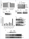
mda-7/IL-24 does not interfere with transcription of Mcl-1. A, top left, DU-145 cells were cotransfected with −203/+10-Mcl-1-pGL2 or pGL2-Basic and pRL-TK-luc plasmids. Cells were incubated for 6 h and then infected with 100 pfu/cell of Ad.vec or Ad.mda-7 for the indicated time, after which the activity of firefly and Renilla luciferase was monitored. Bars, SD (n = 3). Top right, DU-145 cells were treated as above, after which the total RNA was isolated and Mcl-1 mRNA was quantified using real-time PCR. Bars, SD (n = 3). Bottom, DU-145 cells were infected with 100 pfu/cell of Ad.vec or Ad.mda-7 for 2 h and then treated with 1 μg/mL actinomycin D (Act D) for 6 or 12 h, after which cell lysates were prepared and subjected to Western blotting to monitor Mcl-1 protein levels. B, top, DU-145 cells were treated with the indicated amount of MG132 or infected with 100 pfu/cell of Ad.vec or Ad.mda-7 for the designated periods, after which cells were lysed and subjected to immunoprecipitation using anti–Mcl-1 antibodies and then immunoblotted with anti-ubiquitin or Mcl-1 antibodies. Bottom left, DU-145 cells infected with Ad.vec or Ad.mda-7 at 100 pfu/cell were exposed to 1 μmol/L MG132 for the designated periods and subjected to Western blot analysis to monitor Mcl-1 protein levels. Bottom right, DU-145 cells were exposed to 1 μmol/L MG132 or 100 pfu/cell Ad.vec alone, simultaneously treated with 1 μmol/L MG132 and infected with 100 pfu/cell of Ad.mda-7, or infected with Ad.mda-7 (100 pfu/cell) for 6 h before adding 1 μmol/L MG132. After 24 h of treatment, cells were lysed and Western blotting was performed as above. C, DU-145 cells were infected with 100 pfu/cell of Ad.vec or Ad.mda-7 for the indicated times, lysates were collected, and proteasome activity was measured as described in Materials and Methods. Bars, SD (n = 3). D, DU-145 cells were infected with 100 pfu/cell of Ad.vec or Ad.mda-7 for the indicated periods, after which protein lysates were prepared and subjected to Western blotting using the indicated antibodies. E, top, DU-145 cells were infected with 100 pfu/cell of Ad.vec or Ad.mda-7 for 16 h and then cells were labeled with [35S]methionine as described in Materials and Methods and chased for the designated periods (h). At the end of the indicated intervals, cells were lysed and Mcl-1 protein was immunoprecipitated using Mcl-1 antibodies and subjected to SDS-PAGE followed by autoradiography. Bottom, DU-145 cells were infected with 100 pfu/cell of Ad.vec or Ad.mda-7; 30 h later, polysomal fractions were purified from the cell lysates. Polysomal RNA was purified from each fraction, and Northern blotting was done with a Mcl-1 cDNA probe. The numbers represent fraction numbers, with fractions 10 to 20 representing the polysomes.
Figure 7
Figure 7
Mcl-1 is an important regulator of mda-7/IL-24-mediated apoptosis. A, DU-145 cells were infected with 100 pfu/cell of Ad.vec or Ad.mda-7, and protein lysates were prepared at the specified times and subjected to Western blotting using indicated antibodies. B, model for mda-7/IL-24 lethality through induction of an ER stress response and Mcl-1 downregulation.

References

    1. Damber JE, Aus G. Prostate cancer. Lancet. 2008;371:1710–21. - PubMed
    1. Mabjeesh NJ, Zhong H, Simons JW. Gene therapy of prostate cancer: current and future directions. Endocr Relat Cancer. 2002;9:115–39. - PubMed
    1. Jiang H, Lin JJ, Su ZZ, Goldstein NI, Fisher PB. Subtraction hybridization identifies a novel melanoma differentiation associated gene, mda-7, modulated during human melanoma differentiation, growth and progression. Oncogene. 1995;11:2477–86. - PubMed
    1. Pestka S, Krause CD, Sarkar D, Walter MR, Shi Y, Fisher PB. Interleukin-10 and related cytokines and receptors. Annu Rev Immunol. 2004;22:929–79. - PubMed
    1. Gupta P, Su ZZ, Lebedeva IV, et al. mda-7/IL-24: multifunctional cancer-specific apoptosis-inducing cytokine. Pharmacol Ther. 2006;111:596–628. - PMC - PubMed

Publication types

MeSH terms