. 2010 Jun 15;70(12):4922-30.
doi: 10.1158/0008-5472.CAN-10-0095. Epub 2010 May 25.

Similar nucleotide excision repair capacity in melanocytes and melanoma cells

Affiliations

Similar nucleotide excision repair capacity in melanocytes and melanoma cells

Shobhan Gaddameedhi et al. Cancer Res. .

Abstract

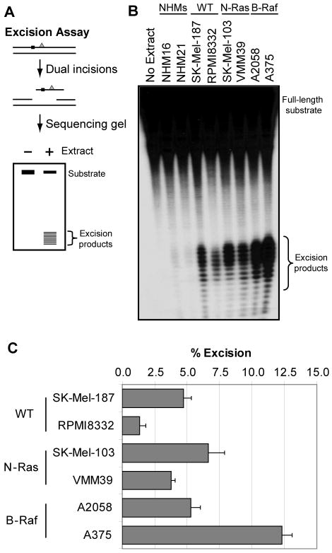
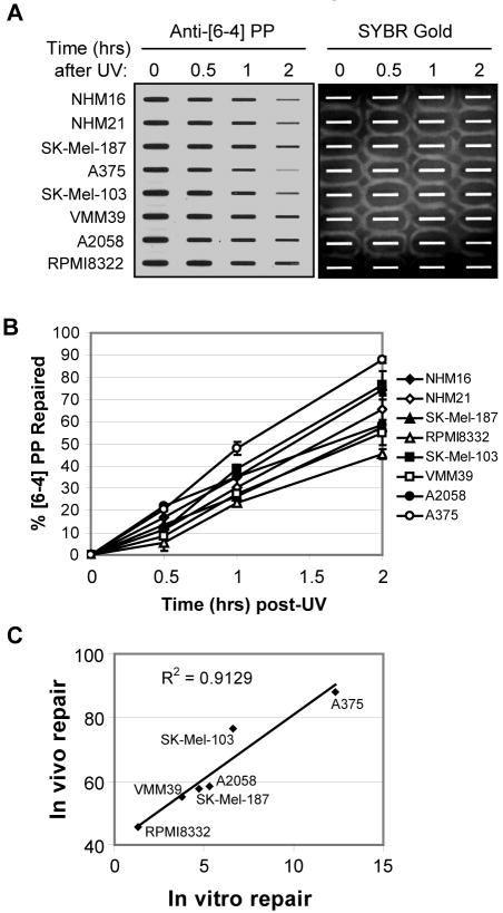
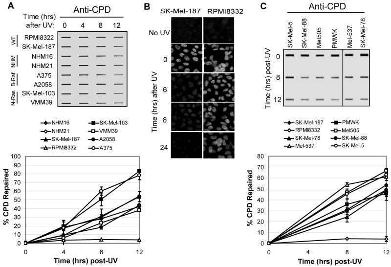
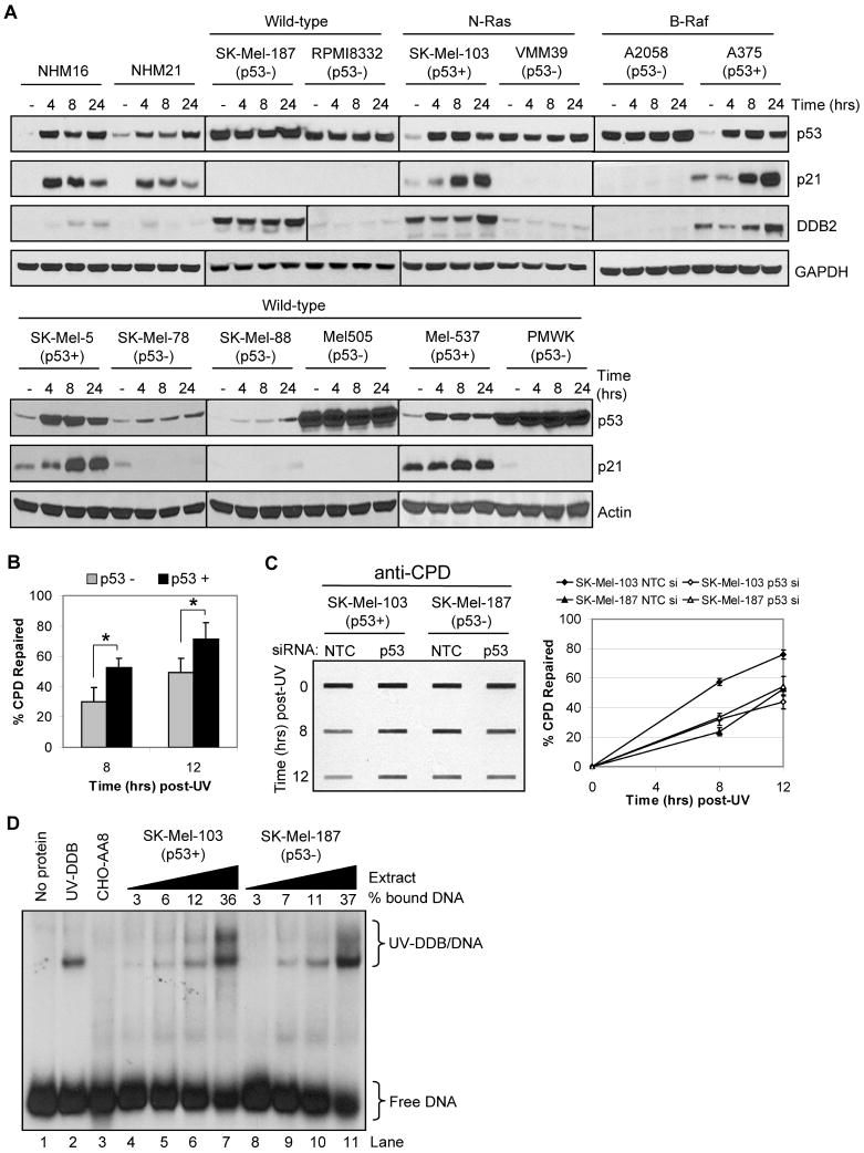
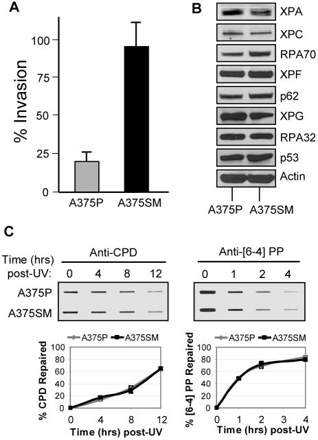
Sunlight UV exposure produces DNA photoproducts in skin that are repaired solely by nucleotide excision repair in humans. A significant fraction of melanomas are thought to result from UV-induced DNA damage that escapes repair; however, little evidence is available about the functional capacity of normal human melanocytes, malignant melanoma cells, and metastatic melanoma cells to repair UV-induced photoproducts in DNA. In this study, we measured nucleotide excision repair in both normal melanocytes and a panel of melanoma cell lines. Our results show that in 11 of 12 melanoma cell lines tested, UV photoproduct repair occurred as efficiently as in primary melanocytes. Importantly, repair capacity was not affected by mutation in the N-RAS or B-RAF oncogenes, nor was a difference observed between a highly metastatic melanoma cell line (A375SM) or its parental line (A375P). Lastly, we found that although p53 status contributed to photoproduct removal efficiency, its role did not seem to be mediated by enhanced expression or activity of DNA binding protein DDB2. We concluded that melanoma cells retain capacity for nucleotide excision repair, the loss of which probably does not commonly contribute to melanoma progression.


Figures

Figure 1
Figure 1. [6-4] photoproduct repair in cell-free extracts
(A) Schematic of the in vitro excision assay. An internally 32P-labeled (black square), 136-bp double-stranded DNA containing a centrally located [6-4] PP (gray triangle) is incubated with cell-free extract for 90 min. The damaged DNA is removed through dual incisions of the DNA at sites bracketing the lesion, resulting in release of 24- to 32-nt-long oligomers (excised products) that are visualized by denaturing PAGE and phosphorimager analysis. (B) Excision repair in cell-free extracts. Cell lines examined included two normal human melanocyte lines (NHMs) and two cell lines each that lack (wild-type: WT) or contain mutations in either N-Ras or B-Raf. (C) Quantitative analyses of excision repair in cell-free extracts. The percent of total radiolabeled material released as excision products represents the amount of excision repair (% excision). Excision assays were performed three times with three independent preparations of cell-free extract for each cell line, and the data indicate the average and standard deviation from these assays.
Figure 2
Figure 2. Immunoslot blot assay for [6-4] photoproduct repair
(A) An immunoslot blot assay was used to measure [6-4] PP repair at various time points post-irradiation in NHMs and melanoma cell lines exposed to 10 J/m2 of UVC. The image shows the [6-4] PP signal detected with an anti-[6-4] PP antibody and SYBR Gold staining to show equal loading of total genomic DNA. (B) Quantitative analysis of the repair assay. The averages and standard deviations are from two independent experiments. (C) Correlation between [6-4] PP repair levels using the immunoslot blot and in vitro cell-free excision assays. For each of the six melanoma cell lines examined, the level of [6-4] PP repair as measured at the two hour time point by immunoslot blot assay (in vivo repair) was plotted along with the amount of [6-4] PP substrate repaired for 90 min in the excision assay in Figure 1 (in vitro repair).
Figure 3
Figure 3. CPD repair in melanocytes and melanoma cell lines
(A) Cells were exposed to UVC (10 J/m2) and harvested at the indicated time points for immunoslot blot analysis of CPD repair. The top image shows a representative experiment. CPD repair assays were performed three times for each cell line and average CPD repair (and standard deviation) graphed below. (B) Immunofluorescence analyses of CPD repair in SK-Mel-187 and RPMI8332 cells. (C) CPD repair in six additional melanoma cell lines with wild-type N-Ras and B-Raf. The graph (right panel) includes SK-Mel-187 and RPMI cells analyzed in (A).
Figure 4
Figure 4. Analysis of p53 and DDB2 functionality and in melanoma cells and melanocytes
(A) Western blot analyses were performed with extracts from the indicated cell lines harvested at various times post UV-irradiation (12.5 J/m2) to monitor p53 functionality, as determined by induction of p21 and DDB2 protein expression. (B) Based on p53 functionality determined in (A), CPD repair measurements for the individual cell lines (Figure 3) were pooled and re-analyzed as a function of p53 status. Data show the average (and standard deviation) of CPD repair for the seven p53 mutant cell lines (RPMI was excluded) and four p53 wild-type cell lines. Asterisks indicate a statistically significant difference (p<0.01; two-tailed Student's t-test) in CPD repair between p53 mutant and wild-type cell lines. (C) Knockdown of p53 in SK-Mel-103 cells inhibits CPD repair. SK-Mel-103 (p53 wild-type) and SK-Mel-187 (p53 mutant) cells were transfected with non-targeting or p53 siRNAs, exposed to UV, and CPD repair measured at the indicated time points. (D) Electrophoretic mobility shift assay of UV-DDB binding to damaged DNA. Cell-free extract from SK-Mel-103 (p53 wild-type) and SK-Mel-187 (p53 mutant) cells were incubated with radiolabeled [6-4] PP substrate DNA and complexes separated on a 5% non-denaturing gel. Purified UV-DDB complex was used as a positive control and CHO-AA8 extract as a negative control. The cell-free extract protein concentrations ranged from 0.5, 1.0, 2.0 or 4.0 μgs per reaction. The experiment was repeated three times, and the average percent binding is indicated.
Figure 5
Figure 5. Excision repair in highly metastatic melanoma cells
(A) Matrigel invasion assay of the highly metastatic cell line (A375SM) compared with its parent cell line (A375P). The results are the averages and standard deviations of three independent experiments. (B) Western blot analyses of the DNA excision repair protein levels of both A375P and A375SM cell lines. (C) Immunoslot blot analysis of CPD and [6-4] PP repair after UV irradiation (10 J/m2) of A375P and A375SM cells. The graphs show the average and standard deviation from two independent experiments.

References

    1. Markovic SN, Erickson LA, Rao RD, et al. Malignant melanoma in the 21st century, part 1: epidemiology, risk factors, screening, prevention, and diagnosis. Mayo Clin Proc. 2007;82:364–80. - PubMed
    1. Gandini S, Sera F, Cattaruzza MS, et al. Meta-analysis of risk factors for cutaneous melanoma: I. Common and atypical naevi. Eur J Cancer. 2005;41:28–44. - PubMed
    1. Ries LA, Wingo PA, Miller DS, et al. The annual report to the nation on the status of cancer, 1973-1997, with a special section on colorectal cancer. Cancer. 2000;88:2398–424. - PubMed
    1. Geller AC, Miller DR, Annas GD, et al. Melanoma incidence and mortality among US whites, 1969-1999. Jama. 2002;288:1719–20. - PubMed
    1. Reardon JT, Sancar A. Nucleotide excision repair. Prog Nucleic Acid Res Mol Biol. 2005;79:183–235. - PubMed

Publication types

MeSH terms