. 2010 Jun 1;70(11):4698-708.
doi: 10.1158/0008-5472.CAN-09-4330. Epub 2010 May 25.

Tetraspanin CD151 regulates growth of mammary epithelial cells in three-dimensional extracellular matrix: implication for mammary ductal carcinoma in situ

Affiliations

Tetraspanin CD151 regulates growth of mammary epithelial cells in three-dimensional extracellular matrix: implication for mammary ductal carcinoma in situ

Vera Novitskaya et al. Cancer Res. .

Abstract

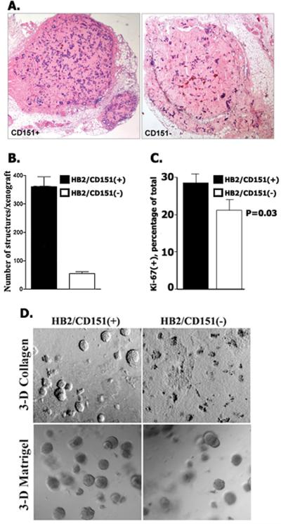
Tetraspanin CD151 is associated with laminin-binding integrins (i.e., alpha(3)beta(1), alpha(6)beta(1), and alpha(6)beta(4)) and regulates tumor cell migration and invasion. Here, we examined the role of CD151 in proliferation of mammary epithelial cells using in vitro and in vivo models. Depletion of CD151 suppressed growth of HB2 cells, a nontumorigenic breast epithelial cell line, in three-dimensional (3D) extracellular matrices (ECM) and in Matrigel-based xenografts. Whereas the presence of alpha(3)beta(1) (but not alpha(6) integrins) was necessary to support growth of HB2 cells in 3D ECM, the pro-proliferative activity of CD151 did not require direct interaction with integrins. Furthermore, depletion of CD151 potentiated formation of the internal lumen and partial restoration of polarity when HB2 cells were cultured in 3D ECM. This correlated with a decrease in phosphorylation levels of extracellular signal-regulated kinase 1/2 and cAkt in CD151-negative cells and increase in activation of caspase-3. Accordingly, the number of CD151-positive colonies with internal lumen was increased by approximately 5-fold when cells were cultured in the presence of MAP/ERK kinase (U0126) and phosphoinositide 3-kinase (LY29004) inhibitors. To establish the physiologic relevance of pro-proliferative and morphogenetic activities of CD151, we analyzed the expression of this tetraspanin in ductal carcinoma in situ (DCIS), which is characterized by neoplastic proliferation of mammary epithelial cells. Strong homogeneous membrane expression of CD151 was found to be associated with a high grade of DCIS (P = 0.004). Taken together, these results strongly suggest that CD151 complexes play a crucial role in the development of hyperproliferative diseases in the mammary gland.


Figures

Figure 1
Figure 1
Expression of CD151 in DCIS and characterization of HB2 xenografts. A. In normal breast the staining is limited to the myoepithelial-basement membrane interface (top left panel, arrows), with sparing of the luminal cells. In DCIS staining was either homogeneously positive (top right panel), with strong membrane staining in all the neoplastic luminal cells, or hetererogeneous (bottom panels), with strong membrane staining of neoplastic cells at the periphery of the duct, but lack of CD151 expression in the cells towards the centre of the ducts, particularly seen as the cells started to form small lumen. B, Cells were injected subcutaneously into nude mice as described in Materials and Methods and xenografts were grown for 8 weeks. Shown morphology (H&E staining) of representative glandular structures.
Figure 2
Figure 2
The effect of CD151 depletion on growth of HB2 cells in vivo and in 3-D ECM. A. Sections of representative HB2/CD151(+) and HB2/CD151(−) xenografts (8 weeks). B. Quantification of structures formed by CD151-positive and CD151-negative HB2 cells in vivo. Shown means of the number of structures. Error bars represent SEM. C. Ki-67 labelling index. Shown results of quantification of Ki-67- immunopositive cells (average from 10 high power fields (x400) ~ 500 cells /sample). D. HB2/CD151(+) and HB2/CD151(−) cells were grown for 10 days in 3-D collagen (top panel) or 3-D Matrigel (bottom panel). Shown representative fields.
Figure 3
Figure 3
The role of CD151-associated integrins in pro-proliferative and morphogenetic activities of the tetraspanin. A. Morphological appearance of colonies formed by indicated HB2 sublines grown for 10 days in 3-D Matrigel. Insets show magnified images of representative colonies. Note, rare colonies formed by HB2/α3β1(−) cells have the appearance of the CD151(+) aggregates. B. Western blots showing expression of CD151 and integrin subunits in cells used in the panel A.
Figure 4
Figure 4
Depletion of CD151 induces lumen formation in colonies formed by HB2 cells in 3-Matrigel. HB2/CD151(+) and HB2/CD151(−) cells were cultured in 3-D Matrigel for 10 days. A. The lumen formation was analysed by the confocal microscopy as described in Materials and Methods. Nuclei were stained with Hoechst 33342. Proportion of the colonies with lumen/partial lumen was quantified in 3 independent experiments with 60-80 colonies assessed in each of the experiments. Error bars represent SEM. B. Activation of Caspase-3 in HB2/CD151(+) and HB2/CD151(−) cells cultured 3-D Matrigel. Colonies were fixed with paraformaldehyde and stained with antibodies to active Caspase-3 (upper panel). Scale bar represents 50μm. Western blot analysis of caspase-3 activation in cultures of HB2/CD151(+) and HB2/CD151(−) cells in 3-D Matrigel (lower panel). C. Depletion of CD151 potentiates detachment-induced activation of Caspase-3. HB2/CD151(+) and HB2/151(−) cells were detached and kept in suspension over poly-HEMA – coated surface in serum-free DMEM for 24 or 48 hours. Proteins were resolved by 12% SDS-PAGE and presence of activated Caspase-3 (and actin) was visualised using Western blotting. Lysates from cells attached to plastic (and incubated in serum-free DMEM) were used as controls (lanes 1 and 2).
Figure 5
Figure 5
Depletion of CD151 causes redistribution of α3β1 integrin and apical relocalisation of Golgi. Cells were grown in Matrigel for 10 days. The colonies were fixed with paraformaldehyde and stained with indicated mAb. In B. actin filaments were visualised using Alexa 594- conjugated phalloidin and nuclei were stained with DAPI. Note fragmented nuclei inside HB2/CD151(−) colony. Shown representative images. Scale bars represent 50μm.
Figure 6
Figure 6
Depletion of CD151 attenuates activation of Erk1/2 and c-Akt in HB2 cells. A. Analysis of signalling pathways in CD151(+) and CD151(−) cells grown in 3-D Matrigel for indicated times. Shown results of one of three independent experiments. B. Inhibition of c-Akt and Erk1/2 signalling pathway suppresses proliferation of HB2/CD151(+) cells in 3-D Matrigel. Cells were cultured in 3-D Matrigel for 10 days in the presence of 10μM U0126 (middle panel) or 10μM LY29004 (bottom panel). Shown images of representative fields. C. HB2 cells were grown in Matrigel for 10 days in the presence of 10μM U0126 (top panel) or 10ηM LY29004 (bottom panel). Nuclei were stained with Hoechst 33342. Shown images of representative fields.

References

    1. Yanez-Mo M, Barreiro O, Gordon-Alonso M, et al. Tetraspanin-enriched microdomains: a functional unit in cell plasma membranes. Trends Cell Biol. 2009;19:434–46. - PubMed
    1. Charrin S, Le NF, Silvie O, et al. Lateral organization of membrane proteins: tetraspanins spin their web. Biochem J. 2009;420:133–54. - PubMed
    1. Hemler ME. Tetraspanin proteins mediate cellular penetration, invasion, and fusion events and define a novel type of membrane microdomain. Annu Rev Cell Dev Biol. 2003;19:397–422. - PubMed
    1. Berditchevski F, Odintsova E. Tetraspanins as regulators of protein trafficking. Traffic. 2007;8:89–96. - PubMed
    1. Odintsova E, Butters TD, Monti E, et al. Gangliosides play an important role in the organisation of CD82-enriched microdomains. Biochem J. 2006;400:315–25. - PMC - PubMed

Publication types

MeSH terms