

In vivo imaging of amyloid deposition in Alzheimer disease using the radioligand 18F-AV-45 (florbetapir [corrected] F 18)
- PMID: 20501908
- PMCID: PMC3101877
- DOI: 10.2967/jnumed.109.069088
In vivo imaging of amyloid deposition in Alzheimer disease using the radioligand 18F-AV-45 (florbetapir [corrected] F 18)
Erratum in
- J Nucl Med. 2010 Aug;51(8):1327
Abstract
An (18)F-labeled PET amyloid-beta (Abeta) imaging agent could facilitate the clinical evaluation of late-life cognitive impairment by providing an objective measure for Alzheimer disease (AD) pathology. Here we present the results of a clinical trial with (E)-4-(2-(6-(2-(2-(2-(18)F-fluoroethoxy)ethoxy)ethoxy)pyridin-3-yl)vinyl)-N-methyl benzenamine ((18)F-AV-45 or florbetapir [corrected] F 18).
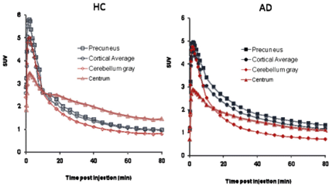
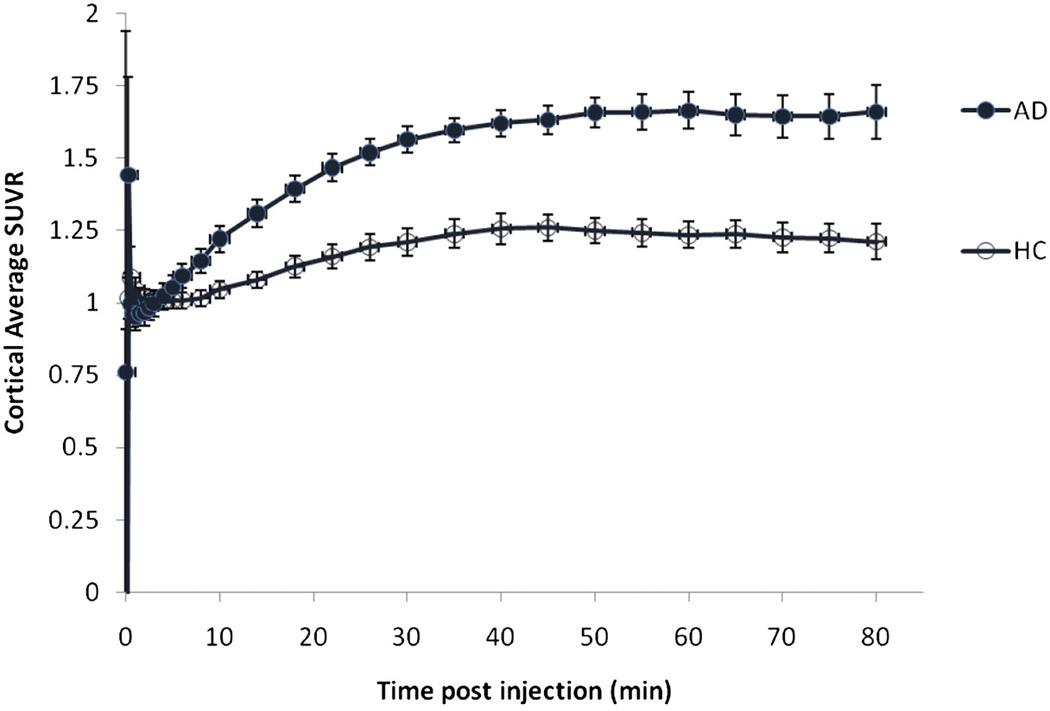
Methods: An open-label, multicenter brain imaging, metabolism, and safety study of (18)F-AV-45 was performed on 16 patients with AD (Mini-Mental State Examination score, 19.3 +/- 3.1; mean age +/- SD, 75.8 +/- 9.2 y) and 16 cognitively healthy controls (HCs) (Mini-Mental State Examination score, 29.8 +/- 0.45; mean age +/- SD, 72.5 +/- 11.6 y). Dynamic PET was performed over a period of approximately 90 min after injection of the tracer (370 MBq [10 mCi]). Standardized uptake values and cortical-to-cerebellum standardized uptake value ratios (SUVRs) were calculated. A simplified reference tissue method was used to generate distribution volume ratio (DVR) parametric maps for a subset of subjects.
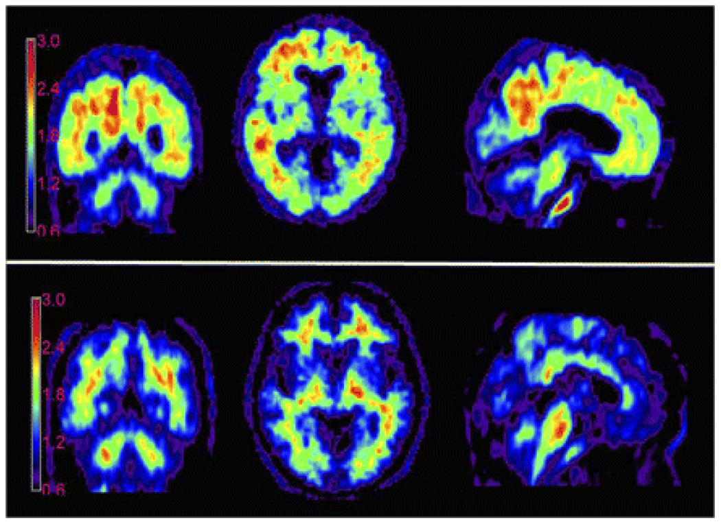
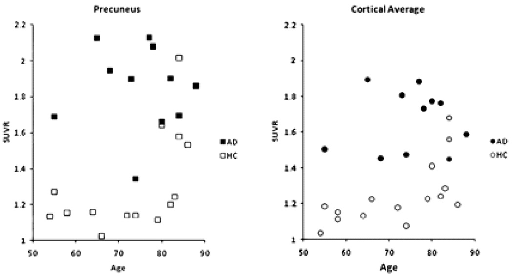
Results: Valid PET data were available for 11 AD patients and 15 HCs. (18)F-AV-45 accumulated in cortical regions expected to be high in Abeta deposition (e.g., precuneus and frontal and temporal cortices) in AD patients; minimal accumulation of the tracer was seen in cortical regions of HCs. The cortical-to-cerebellar SUVRs in AD patients showed continual substantial increases through 30 min after administration, reaching a plateau within 50 min. The 10-min period from 50 to 60 min after administration was taken as a representative sample for further analysis. The cortical average SUVR for this period was 1.67 +/- 0.175 for patients with AD versus 1.25 +/- 0.177 for HCs. Spatially normalized DVRs generated from PET dynamic scans were highly correlated with SUVR (r = 0.58-0.88, P < 0.005) and were significantly greater for AD patients than for HCs in cortical regions but not in subcortical white matter or cerebellar regions. No clinically significant changes in vital signs, electrocardiogram, or laboratory values were observed.
Conclusion: (18)F-AV-45 was well tolerated, and PET showed significant discrimination between AD patients and HCs, using either a parametric reference region method (DVR) or a simplified SUVR calculated from 10 min of scanning 50-60 min after (18)F-AV-45 administration.
Figures
References
-
- Selkoe DJ. Defining molecular targets to prevent Alzheimer disease. Arch Neurol. 2005;62(2):192–195. - PubMed
-
- Hyman BT, Trojanowski JQ. Consensus recommendations for the postmortem diagnosis of Alzheimer disease from the National Institute on Aging and the Reagan Institute Working Group on diagnostic criteria for the neuropathological assessment of Alzheimer disease. J Neuropathol Exp Neurol. 1997;56(10):1095–1097. - PubMed
-
- Mirra SS, Heyman A, McKeel D, et al. The Consortium to Establish a Registry for Alzheimer's Disease (CERAD). Part II. Standardization of the neuropathologic assessment of Alzheimer's disease. Neurology. 1991;41(4):479–486. - PubMed
-
- Hock C, Konietzko U, Streffer JR, et al. Antibodies against beta-amyloid slow cognitive decline in Alzheimer's disease. Neuron. 2003;38(4):547–554. - PubMed
-
- Pike KE, Savage G, Villemagne VL, et al. Beta-amyloid imaging and memory in non-demented individuals: evidence for preclinical Alzheimer's disease. Brain. 2007;130(Pt 11):2837–2844. - PubMed
Publication types
MeSH terms
Substances
Grants and funding
LinkOut - more resources
Full Text Sources
Other Literature Sources
Medical