. 2010 May;22(5):1549-63.
doi: 10.1105/tpc.110.075630. Epub 2010 May 25.

Identification of the 2-hydroxyglutarate and isovaleryl-CoA dehydrogenases as alternative electron donors linking lysine catabolism to the electron transport chain of Arabidopsis mitochondria

Affiliations

Identification of the 2-hydroxyglutarate and isovaleryl-CoA dehydrogenases as alternative electron donors linking lysine catabolism to the electron transport chain of Arabidopsis mitochondria

Wagner L Araújo et al. Plant Cell. 2010 May.

Abstract

The process of dark-induced senescence in plants is relatively poorly understood, but a functional electron-transfer flavoprotein/electron-transfer flavoprotein:ubiquinone oxidoreductase (ETF/ETFQO) complex, which supports respiration during carbon starvation, has recently been identified. Here, we studied the responses of Arabidopsis thaliana mutants deficient in the expression of isovaleryl-CoA dehydrogenase and 2-hydroxyglutarate dehydrogenase to extended darkness and other environmental stresses. Evaluations of the mutant phenotypes following carbon starvation induced by extended darkness identify similarities to those exhibited by mutants of the ETF/ETFQO complex. Metabolic profiling and isotope tracer experimentation revealed that isovaleryl-CoA dehydrogenase is involved in degradation of the branched-chain amino acids, phytol, and Lys, while 2-hydroxyglutarate dehydrogenase is involved exclusively in Lys degradation. These results suggest that isovaleryl-CoA dehydrogenase is the more critical for alternative respiration and that a series of enzymes, including 2-hydroxyglutarate dehydrogenase, plays a role in Lys degradation. Both physiological and metabolic phenotypes of the isovaleryl-CoA dehydrogenase and 2-hydroxyglutarate dehydrogenase mutants were not as severe as those observed for mutants of the ETF/ETFQO complex, indicating some functional redundancy of the enzymes within the process. Our results aid in the elucidation of the pathway of plant Lys catabolism and demonstrate that both isovaleryl-CoA dehydrogenase and 2-hydroxyglutarate dehydrogenase act as electron donors to the ubiquinol pool via an ETF/ETFQO-mediated route.


Figures
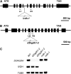
Figure 1.
Figure 1.
A Schematic Representation of the Sites of T-DNA Insertion in the Mutants. (A) and (B) Genomic structure of IVDH (A) and D2HGDH (B). Arrowheads represent positions of primers used for genotyping of wild-type and mutant lines; closed boxes indicate exons. In ivdh-1, the T-DNA is inserted in intron 5 of IVDH. In d2hgdh1-2, the T-DNA is inserted in intron 6 of D2HGDH. (C) RT-PCR analysis on total RNA from the wild type (WT) and the d2hgdh1-2 and ivdh-1 mutant lines, with primer sets for the genes indicated on the left. The positions of the primers in the IVDH and D2HGDH genomic loci are represented in (A) and (B), respectively.
Figure 2.
Figure 2.
Phenotype of Arabidopsis Mutants under Extended Dark Treatment. (A) Images of 4-week-old, short-day-grown Arabidopsis plants immediately (0 d) and after further treatment for 15 d in darkness conditions. The leaves of the ETFQO mutants etfqo-1 and etfqo-2 and of the ivdh-1 mutant were yellowed and dehydrated following 15 d of growth in darkness compared with the wild-type (WT) control (Col-0). Additionally, the complemented lines of each genotype rescue the wild-type phenotype observed under darkness conditions. (B) to (D) Chlorophyll content (B), chlorophyll a/b ratio (C), and Fv/Fm (D), the maximum quantum yield of PSII electron transport, of leaves of 4-week-old, short-day-grown, Arabidopsis plants after further treatment for 0, 3, 7, 10, and 15 d in extended darkness. Values are means ± se of six independent samplings; an asterisk indicates values that were determined by the Student's t test to be significantly different (P < 0.05) from the wild type. FW, fresh weight.
Figure 3.
Figure 3.
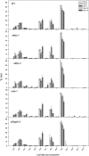
Leaf Fatty Acid Composition in Arabidopsis Mutants under Extended Dark Treatment. Data (in mol %) represent mean ± se for six independent samplings of 9th or 10th leaves of 4-week-old, short-day-grown Arabidopsis plants after treatment for 0, 3, 7, 10, and 15 d in extended darkness. Fatty acid composition was analyzed by GC of fatty acid methyl esters. WT, wild type.
Figure 4.
Figure 4.
Acyl-CoA Profiles in Arabidopsis Mutants under Extended Dark Treatment. Data (in mol %) represent means ± se for six independent samplings of the 9th or 10th leaves of 4-week-old, short-day-grown Arabidopsis plants after further growth for 0, 3, 7, 10, and 15 d in extended darkness. Samples of 10 mg (fresh weight) each were derivatized to their acyl-etheno-CoA esters, separated by HPLC, and detected fluorometrically. WT, wild type.
Figure 5.
Figure 5.
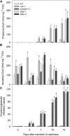
Metabolic Phenotype of Arabidopsis Mutants in Extended Dark Treatment. Phytanoyl-CoA (A), isovaleryl-CoA (B), and 2-hydroxyglutarate (C) profiles in Arabidopsis mutants under extended dark treatment. Samples were taken from leaves of 4-week-old, short-day-grown Arabidopsis plants after treatment for 0, 3, 7, 10, and 15 d in extended darkness. Values are means ± se of six independent samplings; an asterisk indicates values that were determined by the Student's t test to be significantly different (P < 0.05) from the wild type (WT). FW, fresh weight.
Figure 6.
Figure 6.
Relative Levels of Sugars and Organic Acids in Arabidopsis Mutants during Extended Dark Conditions as Measured by GC-MS. The y axis values represent the metabolite level relative to the wild type (WT). Data were normalized to the mean response calculated for the 0-d dark-treated leaves of the wild type. Values presented are means ± se of determinations on six independent samplings; an asterisk indicates values that were determined by the Student's t test to be significantly different (P < 0.05) from the wild type.
Figure 7.
Figure 7.
Relative Levels of Amino Acids in Arabidopsis Mutants during Extended Dark Conditions as Measured by GC-MS. Levels of the indicated amino acids are presented as in Figure 6. WT, wild type.
Figure 8.
Figure 8.
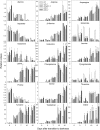
Redistribution of Heavy Label Following Amino Acid Feeding of Arabidopsis Mutant and Wild-Type Leaves. The 9th or 10th leaves of 4-week-old, short-day-grown Arabidopsis plants after treatment for 0, 10, or 15 d in extended darkness were harvested and fed via the petiole with either [U-13C]-Val (A) or [U-13C]-Lys (B) solution. Values represent absolute redistribution of the label and are given as means ± se of determinations on six independent samplings; an asterisk indicates values that were determined by the Student's t test to be significantly different (P < 0.05) from the wild type. FW, fresh weight.
Figure 9.
Figure 9.
Metabolic Model Showing the Involvement of IVDH and D2HGDH in Feeding Electrons to the Mitochondrial Respiration Electron Chain. Isovaleryl-CoA may be produced by BCAA catabolism, chlorophyll degradation, and/or Lys degradation, whereas HG can be produced either in the peroxisome or from the Lys derivative l-pipecolate as in nonplant systems. Thus, the electrons generated are transferred to the respiratory chain through the ubiquinol pool via an ETF/ETFQO system. Dotted arrows represent possible transport processes. 2-OG, 2-oxoglutarate; HG, d-2-hydroxyglutarate; e-, electron; UQ, ubiquinone; 3-MC-CoA, 3-methylcrotonyl-CoA; KAT2, keto-acyl-thiolase 2; chl, chlorophyll; D-2HGS, 2-hydroxyglutarate synthase; 1, conversion of chlorophyll b to a by chlorophyll b reductase.

Similar articles

Cited by

References

    1. Achouri Y., Noel G., Vertommen D., Rider M.H., Veiga-Da-Cunha M., van Schaftingen E. (2004). Identification of a dehydrogenase acting on D-2-hydroxyglutarate. Biochem. J. 381: 35–42 - PMC - PubMed
    1. Angelovici R., Fait A., Zhu X., Szymanski J., Feldmesser E., Fernie A.R., Galili G. (2009). Deciphering transcriptional and metabolic networks associated with lysine metabolism during Arabidopsis seed development. Plant Physiol. 151: 2058–2072 - PMC - PubMed
    1. Azevedo R.A., Arruda P. (April 7, 2010). High-lysine maize: the key discoveries that have made it possible. Amino Acids, http://dx.doi.org/10.1007/s00726-010-0576-5 - DOI - PubMed
    1. Azevedo R.A., Lea P.J. (2001). Lysine metabolism in higher plants. Amino Acids 20: 261–279 - PubMed
    1. Beckmann J.D., Frerman F.E. (1985). Electron-transfer flavoprotein:ubiquinone oxidoreductase from pig liver: Purification and molecular, redox, and catalytic properties. Biochemistry 24: 3913–3921 - PubMed

Publication types

MeSH terms