. 2010 May 25;3(123):ra40.
doi: 10.1126/scisignal.2000727.

The balance of WNT and FGF signaling influences mesenchymal stem cell fate during skeletal development

Affiliations

The balance of WNT and FGF signaling influences mesenchymal stem cell fate during skeletal development

Takamitsu Maruyama et al. Sci Signal. .

Abstract

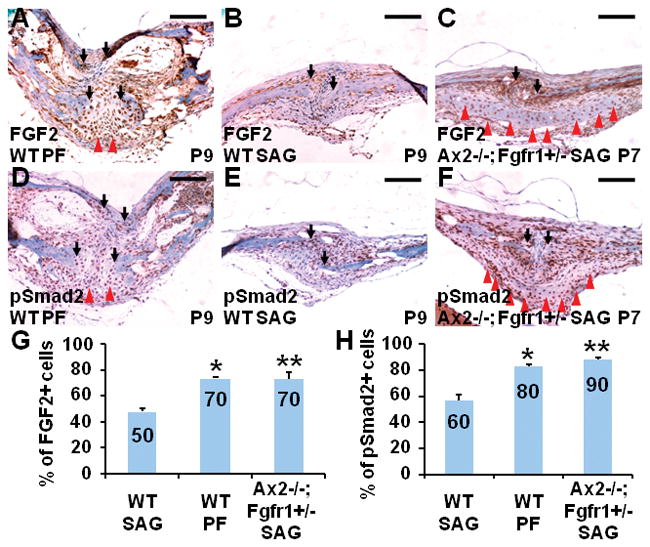
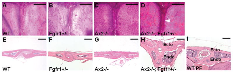
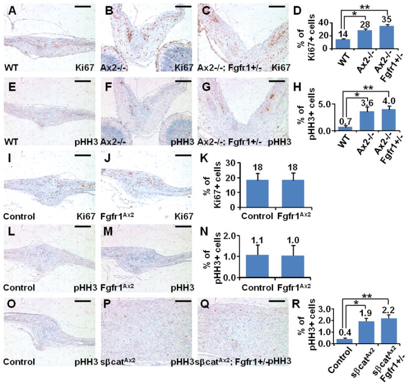
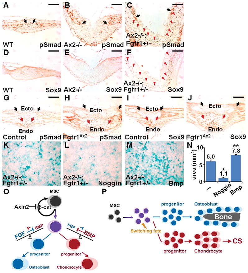
Craniosynostosis, a developmental disorder resulting from premature closure of the gaps (sutures) between skull bones, can be caused by excessive intramembranous ossification, a type of bone formation that does not involve formation of a cartilage template (chondrogenesis). Here, we show that endochondral ossification, a type of bone formation that proceeds through a cartilage intermediate, caused by switching the fate of mesenchymal stem cells to chondrocytes, can also result in craniosynostosis. Simultaneous knockout of Axin2, a negative regulator of the WNT-beta-catenin pathway, and decreased activity of fibroblast growth factor (FGF) receptor 1 (FGFR1) in mice induced ectopic chondrogenesis, leading to abnormal suture morphogenesis and fusion. Genetic analyses revealed that activation of beta-catenin cooperated with FGFR1 to alter the lineage commitment of mesenchymal stem cells to differentiate into chondrocytes, from which cartilage is formed. We showed that the WNT-beta-catenin pathway directly controlled the stem cell population by regulating its renewal and proliferation, and indirectly modulated lineage specification by setting the balance of the FGF and bone morphogenetic protein pathways. This study identifies endochondral ossification as a mechanism of suture closure during development and implicates this process in craniosynostosis.


Conflict of interest statement

Competing interests: Mice reported in this paper are available, but require a material transfer agreement from the University of Rochester.

Figures

Fig. 1
Fig. 1
Ectopic chondrogenesis caused by loss of Axin2 and decreased FGFR1 induces ectopic chondrogenesis. (A to D, A′ to D′) Skeletal staining of the P7 skulls with AB shows that ectopic chondrogenesis (arrow) occurs in the SAG suture of Ax2−/−; Fgfr1+/− (100%, n = 6) but not in the SAG suture of wild-type (WT; 100%, n = 4) or in Fgfr1+/− (100%, n = 4) mice. An asterisk indicates faint AB reactivity observed in a few Ax2−/− (37%, n = 11). (E to H, E′ to H′) AB staining, (I to L) immunostaining of Sox9, and (M to P) immunostaining of type II collagen (Col II) reveals that combined loss of Axin2 and decreased FGFR1 increases the number of chondrogenic precursors and chondrocytes in the SAG suture at P7. Sections were counterstained by eosin in red. Scale bars, 2 mm (A to D); 1 mm (A′ to D′); 200 μm (E to H); and 50 μm (E′ to H′ and I to P).
Fig. 2
Fig. 2
Increased abundance of FGF2 and phosphorylated Smad2 coincides with development of the normal PF suture and the mutant SAG suture that exhibits a PF suture–like structure. Sutures of P9 WT and double-mutant mice were immunostained for FGF2 (A to C) and pSmad2 (D to F). Brown color is positive for FGF2 or pSmad2 and blue indicates hematoxylin counterstaining. Black arrows indicate the osteogenic fronts, which are positive for FGF2 and pSmad2 in WT mice during PF and SAG suture morphogenesis. The areas surrounding the bone plates are also positive. In addition to these intramembranous ossification regions, red arrowheads indicate the endocranial layer of the PF suture mesenchyme that is undergoing chondrogenesis, which is also positive for FGF2 and pSmad2 in the WT mice. The PF-like SAG suture in the Ax2−/−; Fgfr1+/− double mutants also exhibited staining for FGF2 and pSmad2 (C and F). In the wild type, the PF suture contains higher percentage of FGF2 (G; *P < 0.001) and pSmad2 positive cells (H; *P < 0.002). The double-mutant SAG suture displays enhanced numbers of cells positive for FGF2 (G; **P < 0.008; n = 3) and pSmad2 (H, **P < 0.001; n = 3). Scale bars, 100 μm.
Fig. 3
Fig. 3
Craniosynostosis occurs in the SAG suture of Ax2−/−; Fgfr1+/− at P50. (A to D) Ventral view of the Alizarin red–stained skulls reveals the closure of the SAG suture (arrowhead) in the Ax2−/−; Fgfr1+/− mutant (73%, n = 11), but not in the WT (100%, n = 3), Fgfr1+/− (100%, n = 4), or Ax2−/− (100%, n = 5). (E to H) Histology of the SAG suture identifies both ectocranial and endocranial layers in the Ax2−/−; Fgfr1+/− mutants, but these two layers are absent in the WT, Fgfr1+/−, and Ax2−/− SAG sutures. The endocranial layer is fused in the Ax2−/−; Fgfr1+/− mutants. (I) This abnormal structure resembles the WT PF suture, which normally is fused at this stage. Endo, endocranial layer; Ecto, ectocranial layer. Scale bars, 1 mm (A to D); 200 μm (E to I).
Fig. 4
Fig. 4
Progression of ectopic chondrogenesis to synostosis is mediated by endochondral ossification in the Ax2−/−; Fgfr1+/− SAG suture. Time course study of endochondral ossification is analyzed by AB, TUNEL, immunostaining of laminin, alkaline phosphatase (ALP) activity, and histology at the indicated stages. (A to C) The asterisk indicates the ectopic chondrocytes found at P10 (A), which are reduced at P15 (B) and P20 (C). (D to F) White arrowheads identify apoptotic cells present during resorption of chondrocytes at P10 (D), which are not present at P15 (E) and P20 (F). (G to I) White arrows indicate the laminin-positive endothelial cells present in the P15 (H) and P20 (I), which are absent in the P10 (G) SAG suture. (J to O) Black arrowheads indicate the site of increasing osteoblastogenesis revealed by ALP staining (J to L), which is accompanied by ectopic bone formation indicated by black arrows (L and O) in histology preparations (M to O). OF, osteogenic front; EC, ectopic chondrocyte; SS, sagittal sinus. Scale bars, 100 μm.
Fig. 5
Fig. 5
Decreased expression of Axin2 precedes chondrogenesis during PF suture fusion. (A to D) Axin2GFP mice, which are positive for GFP in Axin2-expressing cells, were used to examine the spatiotemporal-specific expression of Axin2 from P5 to P28. Whole-mount GFP analysis reveals that Axin2 is expressed in the frontal suture before P5 (A, arrow), but diminished (arrowheads) at P7 (B), P9 (C), and P28 (D). In contrast, Axin2 is consistently present in other sutures, including SAG, coronal, and lambdoid sutures, at postnatal stages. (E to H) Chondrogenesis and down-regulation of Axin2 are coordinated in the developing PF suture. (I to L) Strong expression of Axin2 and no AB-stained chondrocytes are found in both P5 and P9 SAG sutures. GFP staining was counterstained by DAPI (4′,6-diamidino-2-phenylindole) in blue. (M to O) β-Catenin activity increases as Axin2 expression diminishes, as determined by nuclear localization of activated β-catenin (ABC) in the PF suture. (P to R) Diminishing Axin2 expression corresponds with induction of chondrocyte progenitors in the PF suture, detected by immunostaining for SOX9. (S to U) The activated β-catenin colocalizes with SOX9 in the skeletal precursors of PF suture at P7. (V to X) The abundance of FGFR1 in the PF suture is also enhanced upon chondrogenesis at P5 to P9. (Y) Statistical analysis of the percentage of cells positive for Axin2, ABC, SOX9, and FGFR1 (data represent the mean ± SEM from three animals). (Z) A diagram illustrates the temporal-specific regulation of Axin2, activated β-catenin, SOX9, and FGFR1, as well as chondrogenesis (represented as intensity of AB) in the developing PF suture. Endo, endocranial layer; Ecto, ectocranial layer; Br, brain. Scale bar, 2 mm (A to D), 100 μm (E to L), 50 μm (M to X).
Fig. 6
Fig. 6
Targeted disruption of Fgfr1 in the Axin2-expressing skeletal precursors accelerates chondrogenesis in the PF suture. (A) A diagram illustrates ablation of Fgfr1 with a system combining tetracycline-dependent activation and Cre-mediated recombination. The transcription factor rtTA is expressed under control of the Axin2 promoter (Ax2 P). In the presence of doxycycline (Dox), rtTA drives the expression of Cre resulting in conditional deletion of Fgfr1 in the Axin2-expressing cells. In the presence of the R26R allele, the Cre-mediated recombination can be monitored by β-Gal staining. (B and C) β-Gal staining of the Fgfr1Ax2; R26R (Axin2-rtTA; TRE-Cre; Fgfr1Fx/Fx; R26R) and control (TRE-Cre; Fgfr1Fx/Fx; R26R) P7 skulls demonstrates the efficacy of Fgfr1 deletion. (D to G) Sections of the β-Gal–stained skull further show that the Cre-mediated recombination occurs in the P7 PF and SAG suture mesenchyme, osteogenic fronts, and periosteum. (H to K) Immunostaining of FGFR1 indicates that its abundance was reduced in the developing PF and SAG suture of Fgfr1Ax2; R26R at P7. The stained sections in brown were counterstained by hematoxylin in blue. (L to O) AB staining reveals that, in the Fgfr1Ax2; R26R mutant, chondrogenesis was accelerated in the PF suture, whereas no effect was detected in the SAG suture at P7 (n = 4). Scale bar, 4 mm (B and C); 100 μm (D to G); 50 μm (H to O).
Fig. 7
Fig. 7
Together, β-catenin and FGF signaling control induction of the chondrocyte fate. In vitro mesenchymal cultures show accelerated chondrogenesis in the Ax2−/−; Fgfr1+/− mutant. (A and B) Primary skeletal precursors, isolated from the WT and Ax2−/−; Fgfr1+/− SAG sutures and parietal bones, were cultured in differentiation media for 3 weeks and chondrocytes were identified by AB staining. (C) Analysis of the stained area shows an ~18-fold increase in the area positive for AB in the Ax2−/−; Fgfr1+/− cultures compared to that of WT cultures. (D to K) Chondrogenesis is affected by stimulation of WNT signaling (with BIO) and inhibition of FGF signaling (with SU5402). Primary skeletal precursors, isolated from skulls (D to G) or bone marrow (H to K), were induced for differentiation in culture with media only (D and H) or with the indicated inhibitors. Graphs show the quantification of the stained area (*P < 0.022; n = 3 individual experiments, data plotted are the mean ± SEM).
Fig. 8
Fig. 8
Expansion of skeletal precursors is affected by increased β-catenin signaling, but not by reduced FGF signaling. Sections of the SAG sutures from animals with the indicated genotypes were immunostained for Ki67 or for phosphorylated histone H3 (pHH3) and quantified. (A to H) Haploid deficiency of Fgfr1 has no effect on the enhanced proliferation of skeletal precursors caused by Axin2 deficiency. The mean percentage of Ki67- or pHH3-positive cells was significantly increased in the Ax2−/− (*P < 0.004, n = 4 for Ki67; P < 0.03; n = 3 for pHH3) and the Ax2−/−; Fgfr1+/− (**P < 0.0006, n = 4 for Ki67; P < 0.009; n = 3 for pHH3), compared to the WT. (I to N) Targeted disruption of Fgfr1 does not alter the skeletal precursor proliferation. There was no significant difference in the number of Ki67- or pHH3-positive cells in the control (TRE-Cre; Fgfr1Fx/Fx) and Fgfr1Ax2 (Axin2-rtTA; TRE-Cre; Fgfr1Fx/Fx) SAG sutures at P0 (n = 3). (O to R) Stimulated proliferation of skeletal precursors in the sβcatAx2 SAG suture is not affected by Fgfr1 haploid deficiency. At P0, regardless of the Fgfr1 genetic background, compared to WT the percentage of pHH3-positive cells was increased in the SAG sutures of mice with mutant β-catenin, both the sβcatAx2 (*P < 0.007; n = 3) and the sβcatAx2; Fgfr1+/− (**P < 0.007; n = 3) mice.
Fig. 9
Fig. 9
BMP signaling is crucial for development of suture mesenchymal stem cells. (A to F) Activation of the BMP pathway and ectopic chondrogenesis coincide in the Ax2−/−; Fgfr1+/− mutants. BMP signaling was detected by immunostaining for phosphorylated SMAD1/5/8, and chondrogenic cells were detected by immunostaining for SOX9 in sections of SAG sutures from P0 animals. Black arrows indicate the area surrounding the osteogenic fronts that contains cells that are positive for pSMAD1/5/8. Red arrowheads indicate that pSMAD1/5/8 is detected in the SOX9-positive chondrogenic progenitors found only in the Ax2−/−; Fgfr1+/− SAG suture undergoing ectopic chondrogenesis. (G to J) BMP signaling is increased in the Fgfr1Ax2 PF suture, which displays accelerated chondrogenesis. Sections of the P7 control (TRE-Cre; Fgfr1Fx/Fx) and Fgfr1Ax2 (Axin2-rtTA; TRE-Cre; Fgfr1Fx/Fx) PF sutures were immunostained for pSMAD1/5/8 and SOX9. The chondrogenic domain with active BMP signaling, defined by the presence of SOX9 and pSMAD1/5/8, was expanded in the Fgfr1Ax2 PF suture compared to the control. Black arrows and red arrows indicate the osteogenic fronts and the chondrogenic areas of the PF suture, respectively. Endo, endocranial layer; Ecto, ectocranial layer. (K to N) BMP signaling is required for chondrocyte differentiation induced by altered β-catenin and FGF signaling. Primary skeletal precursors, isolated from the Ax2−/−; Fgfr1+/− calvaria, were cultured in differentiation media with or without BMP or Noggin for 3 weeks. Quantification of the ABstained area shows that Noggin significantly suppressed chondrogenesis (*P < 0.005; n = 3 individual experiments, mean ± SEM) and BMP enhanced (**P < 0.028; n = 3 individual experiments, mean ± SEM) chondrogenesis. (O) Diagram illustrates the model for development of mesenchymal stem cells in calvarial morphogenesis. (P) Diagram illustrates endochondral ossification caused by switching the fate of mesenchymal stem cells resulting in craniosynostosis (CS). Scale bar, 100 μm (A to J).

Similar articles

Cited by

References

    1. Wilkie AO, Morriss-Kay GM. Genetics of craniofacial development and malformation. Nat Rev Genet. 2001;2:458–468. - PubMed
    1. Cohen MM, MacLean RE. Craniosynostosis: Diagnosis, Evaluation, and Management. Oxford University Press; New York: 2000. p. 454.
    1. Opperman LA. Cranial sutures as intramembranous bone growth sites. Dev Dyn. 2000;219:472–485. - PubMed
    1. Hall BK. Bone. Telford Press; Caldwell, NJ: 1990. p. v.
    1. Wilkie AO. Craniosynostosis: Genes and mechanisms. Hum Mol Genet. 1997;6:1647–1656. - PubMed

Publication types

MeSH terms

LinkOut - more resources