. 2010 Apr 6:6:32.
doi: 10.3762/bjoc.6.32.

Molecular recognition of organic ammonium ions in solution using synthetic receptors

Affiliations

Molecular recognition of organic ammonium ions in solution using synthetic receptors

Andreas Späth et al. Beilstein J Org Chem. .

Abstract

Ammonium ions are ubiquitous in chemistry and molecular biology. Considerable efforts have been undertaken to develop synthetic receptors for their selective molecular recognition. The type of host compounds for organic ammonium ion binding span a wide range from crown ethers to calixarenes to metal complexes. Typical intermolecular interactions are hydrogen bonds, electrostatic and cation-π interactions, hydrophobic interactions or reversible covalent bond formation. In this review we discuss the different classes of synthetic receptors for organic ammonium ion recognition and illustrate the scope and limitations of each class with selected examples from the recent literature. The molecular recognition of ammonium ions in amino acids is included and the enantioselective binding of chiral ammonium ions by synthetic receptors is also covered. In our conclusion we compare the strengths and weaknesses of the different types of ammonium ion receptors which may help to select the best approach for specific applications.

Keywords: amino acids; ammonium ion; molecular recognition; synthetic receptors.


Figures

Figure 1
Figure 1
Biologically important amines and quaternary ammonium salts: histamine (1), dopamine (2) and acetylcholine (3).
Figure 2
Figure 2
Crown ether 18-crown-6.
Figure 3
Figure 3
Conformations of 18-crown-6 (4) in solvents of different polarity.
Figure 4
Figure 4
Binding topologies of the ammonium ion depending on the crown ring size.
Figure 5
Figure 5
A “pseudorotaxane” structure consisting of 24-crown-8 and a secondary ammonium ion (5); R = Ph.
Figure 6
Figure 6
Typical examples of azacrown ethers, cryptands and related aza macrocycles.
Figure 7
Figure 7
Binding of ammonium to azacrown ethers and cryptands [–113].
Figure 8
Figure 8
A 19-crown-6-ether with decalino blocking groups (11) and a thiazole-dibenzo-18-crown-6-ether (12).
Figure 9
Figure 9
1,3-Bis(6-oxopyridazin-1-yl)propane derivatives 13 and 14 by Campayo et al.
Figure 10
Figure 10
Fluorescent azacrown-PET-sensors based on coumarin.
Figure 11
Figure 11
Two different pyridino-cryptands (17 and 18) compared to a pyridino-crown (19); chiral ammonium ions as guests (20a–c).
Figure 12
Figure 12
Pyridino-18-crown-6 ligand (21), a similar acridino-18-crown-6 ligand (22) and a structurally related bispyridyl (bpy)-18-crown-6 receptor 23.
Figure 13
Figure 13
Ciral pyridine-azacrown ether receptors 24.
Figure 14
Figure 14
Chiral 15-crown-5 receptors 26 and an analogue 18-crown-6 ligand 27 derived from amino alcohols.
Figure 15
Figure 15
C2-symmetric chiral 18-crown-6 amino alcohol derivatives 28 and related macrocycles.
Figure 16
Figure 16
Macrocycles with diamide-diester groups (30).
Figure 17
Figure 17
C2-symmetric chiral aza-18-crown-6 ethers (31) with phenethylamine residues.
Figure 18
Figure 18
Chiral C-pivot p-methoxy-phenoxy-lariat ethers.
Figure 19
Figure 19
Chiral lariat crown ether 34.
Figure 20
Figure 20
Sucrose-based chiral crown ether receptors 36.
Figure 21
Figure 21
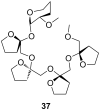
Permethylated fructooligosaccharide 37 showing induced-fit chiral recognition.
Figure 22
Figure 22
Biphenanthryl-18-crown-6 derivative 38.
Figure 23
Figure 23
Chiral lariat crown ethers derived from binol by Fuji et al.
Figure 24
Figure 24
Chiral phenolic crown ether 41 with “aryl chiral barriers” and guest amines.
Figure 25
Figure 25
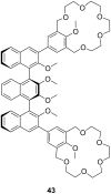
Chiral bis-crown receptor 43 with a meso-ternaphthalene backbone.
Figure 26
Figure 26
Chromogenic pH-dependent bis-crown chemosensor 44 for diamines.
Figure 27
Figure 27
Triamine guests for binding to receptor 44.
Figure 28
Figure 28
Chiral bis-crown phenolphthalein chemosensors 46.
Figure 29
Figure 29
Crown ether amino acid 47.
Figure 30
Figure 30
Luminescent receptor 48 for bis-alkylammonium guests.
Figure 31
Figure 31
Luminescent CEAA (49a), a bis-CEAA receptor for amino acids (49b) and the structure of lysine binding.
Figure 32
Figure 32
Luminescent CEAA tripeptide for binding small peptides.
Figure 33
Figure 33
Bis crown ether 51a self assembles co-operatively with C60-ammonium ion 51b.
Figure 34
Figure 34
Triptycene-based macrotricyclic dibenzo-[24]-crown-8 ether host 52 and guests.
Figure 35
Figure 35
Copper imido diacetic acid azacrown receptor 53a and the suggested His-Lys binding motif; a copper imido triacetic acid azacrown receptor 53b and the target binding area (R = COO, CONHCH2COO, CONHCH2COOCH3, CONHCH2CONHCH2CONH2; R′ = H, CH3,CH2-CH(CH3)2, CH2CH2CONH2).
Figure 36
Figure 36
Urea (54) and thiourea (55) benzo crown receptor for transport and extraction of amino acids.
Figure 37
Figure 37
Crown pyryliums ion receptors 56 for amino acids.
Figure 38
Figure 38
Ditopic sulfonamide bridged crown ether receptor 57.
Figure 39
Figure 39
Luminescent peptide receptor 58.
Figure 40
Figure 40
Luminescent receptor 59 for the detection of D-glucosamine hydrochloride in water/ethanol and luminescent receptor 60 for ω-amino acids.
Figure 41
Figure 41
Guanidinium azacrown receptor 61 for simple amino acids and ditopic receptor 62 with crown ether and polyammonium macrocycle for GABA binding.
Figure 42
Figure 42
Chiral bicyclic guanidinium azacrown receptor 63 and similar receptor 64 for the enantioselective transport of simple amino acids into organic phases.
Figure 43
Figure 43
Receptors for zwitterionic species based on luminescent CEAAs.
Figure 44
Figure 44
1,10-Azacrown ethers with sugar podand arms and the anticancer agent busulfan.
Figure 45
Figure 45
Benzo-18-crown-6 modified β-cyclodextrin 69 and β-cyclodextrin functionalized with diaza-18-crown-6 at primary face (70).
Figure 46
Figure 46
Receptors for colorimetric detection of primary and secondary ammonium ions.
Figure 47
Figure 47
Porphyrine-crown-receptors 72.
Figure 48
Figure 48
Porphyrin-crown ether conjugate 73 and fullerene-ammonium ion guest 74.
Figure 49
Figure 49
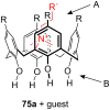
Calix[4]arene (75a), homooxocalix[4]arene (75b) and resorcin[4]arene (75c) compared (R = H, alkyl chain).
Figure 50
Figure 50
Calix[4]arene and ammonium ion guest (R = H, alkyl, OAcyl etc.), possible binding sites; A: co-ordination of cationic or neutral guests (cation–π-interaction), B: binding site for cationic guests (ion–dipole-interaction or H-bonding).
Figure 51
Figure 51
Typical guests for studies with calixarenes and related molecules.
Figure 52
Figure 52
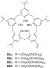
Lower rim modified p-tert-butylcalix[5]arenes 82.
Figure 53
Figure 53
The first example of a water soluble calixarene.
Figure 54
Figure 54
Sulfonated water soluble calix[n]arenes that bind ammonium ions.
Figure 55
Figure 55
Displacement assay for acetylcholine (3) with a sulfonato-calix[6]arene (84b).
Figure 56
Figure 56
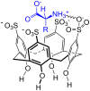
Amino acid inclusion in p-sulfonatocalix[4]arene (84a).
Figure 57
Figure 57
Calixarene receptor family 86 with upper and lower rim functionalization.
Figure 58
Figure 58
Calix[6]arenes 87 with one carboxylic acid functionality.
Figure 59
Figure 59
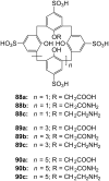
Sulfonated calix[n]arenes with mono-substitution at the lower rim systematically studied on their response to amino acids.
Figure 60
Figure 60
Cyclotetrachromotropylene host (91) and its binding to lysine (81c).
Figure 61
Figure 61
Calixarenes 92 and 93 with phosphonic acids groups.
Figure 62
Figure 62
Calix[4]arene tetraphosphonic acid (94a) and a double bridged analogue (94b).
Figure 63
Figure 63
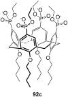
Calix[4]arene tetraphosphonic acid ester (92c) for surface recognition experiments.
Figure 64
Figure 64
Calixarene receptors 95 with α-aminophosphonate groups.
Figure 65
Figure 65
A bridged homocalix[3]arene 95 and a distally bridged homocalix[4]crown 96.
Figure 66
Figure 66
Homocalix[3]arene ammonium ion receptor 97a and the Reichardt’s dye (97b) for colorimetric assays.
Figure 67
Figure 67
Chromogenic diazo-bridged calix[4]arene 98.
Figure 68
Figure 68
Calixarene receptor 99 by Huang et al.
Figure 69
Figure 69
Calixarenes 100 reported by Parisi et al.
Figure 70
Figure 70
Guest molecules for inclusion in calixarenes 100: DAP × 2 HCl (101a), APA (101b) and Lys-OMe × 2 HCl (101c).
Figure 71
Figure 71
Different N-linked peptido-calixarenes open and with glycol chain bridges.
Figure 72
Figure 72
(S)-1,1′-Bi-2-naphthol calixarene derivative 104 published by Kubo et al.
Figure 73
Figure 73
A chiral ammonium-ion receptor 105 based on the calix[4]arene skeleton.
Figure 74
Figure 74
R-/S-phenylalaninol functionalized calix[6]arenes 106a and 106b.
Figure 75
Figure 75
Capped homocalix[3]arene ammonium ion receptor 107.
Figure 76
Figure 76
Two C3 symmetric capped calix[6]arenes 108 and 109.
Figure 77
Figure 77
Phosphorous-containing rigidified calix[6]arene 110.
Figure 78
Figure 78
Calix[6]azacryptand 111.
Figure 79
Figure 79
Further substituted calix[6]azacryptands 112.
Figure 80
Figure 80
Resorcin[4]arene (75c) and the cavitands (113).
Figure 81
Figure 81
Tetrasulfonatomethylcalix[4]resorcinarene (114).
Figure 82
Figure 82
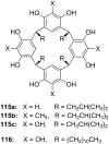
Resorcin[4]arenes (115a/b) and pyrogallo[4]arenes (115c, 116).
Figure 83
Figure 83
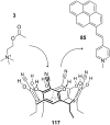
Displacement assay for acetylcholine (3) with tetracyanoresorcin[4]arene (117).
Figure 84
Figure 84
Tetramethoxy resorcinarene mono-crown-5 (118).
Figure 85
Figure 85
Components of a resorcinarene based displacement assay for ammonium ions.
Figure 86
Figure 86
Chiral basket resorcin[4]arenas 121.
Figure 87
Figure 87
Resorcinarenes with deeper cavitand structure (122).
Figure 88
Figure 88
Resorcinarene with partially open deeper cavitand structure (123).
Figure 89
Figure 89
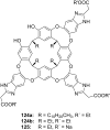
Water-stabilized deep cavitands with partially structure (124, 125).
Figure 90
Figure 90
Charged cavitands 126 for tetralkylammonium ions.
Figure 91
Figure 91
Ditopic calix[4]arene receptor 127 capped with glycol chains.
Figure 92
Figure 92
A calix[5]arene dimer for diammonium salt recognition.
Figure 93
Figure 93
Calixarene parts 92c and 129 for the formation molecular capsules.
Figure 94
Figure 94
Encapsulation of a quaternary ammonium cation by two resorcin[4]arene molecules (NMe4+@[75c]2 × Cl × 6MeOH × H2O; for clarity, solvent molecules and counterions have been omitted).
Figure 95
Figure 95
Encapsulation of a quaternary ammonium cation by six resorcin[4]arene molecules (NMe3D+@[130]6 × Cl; solvent molecules, substituents and counterions are omitted for clarity; the last two resorcinarene calixes are arranged behind and in front of the scheme’s plane).
Figure 96
Figure 96
Structure and schematic of cucurbit[6]uril (CB[6], 131a).
Figure 97
Figure 97
Cyclohexanocucurbit[6]uril (CB′[6], 132) and the guest molecule spermine (133).
Figure 98
Figure 98
α,α,δ,δ-Tetramethylcucurbit[6]uril (134).
Figure 99
Figure 99
Structure of the cucurbituril-phthalhydrazide analogue 135.
Figure 100
Figure 100
Organic cavities for the displacement assay for amine differentiation.
Figure 101
Figure 101
Displacement assay methodology for diammonium- and related guests involving cucurbiturils and some guests.
Figure 102
Figure 102
Nor-seco-Cucurbituril (±)-bis-ns-CB[6] (140) and guest molecules.
Figure 103
Figure 103
The cucurbit[6]uril based complexes 141 for chiral discrimination.
Figure 104
Figure 104
Cucurbit[7]uril (131c) and its ferrocene guests (142) opposed.
Figure 105
Figure 105
Cucurbit[7]uril (131c) guest inclusion and representative guests.
Figure 106
Figure 106
Cucurbit[7]uril (131c) binding to succinylcholine (145) and different bis-ammonium and bis-phosphonium guests.
Figure 107
Figure 107
Paraquat-cucurbit[8]uril complex 149.
Figure 108
Figure 108
Gluconuril-based ammonium receptors 150.
Figure 109
Figure 109
Examples of clefts (151a), tweezers (151b, 151c, 151d) and clips (151e).
Figure 110
Figure 110
Kemp’s triacid (152a), on example of Rebek’s receptors (152b) and guests.
Figure 111
Figure 111
Amino acid receptor (154) by Rebek et al.
Figure 112
Figure 112
Hexagonal lattice designed hosts by Bell et al.
Figure 113
Figure 113
Bell’s amidinium receptor (156) and the amidinium ion (157).
Figure 114
Figure 114
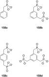
Aromatic phosphonic acids.
Figure 115
Figure 115
Xylene phosphonates 159 and 160a/b for recognition of amines and amino alcohols.
Figure 116
Figure 116
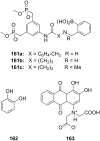
Bisphosphonate recognition motif 161 for a colorimetric assay with alizarin complexone (163) for catechols (162).
Figure 117
Figure 117
Bisphosphonate/phosphate clip 164 and bisphosphonate cleft 165.
Figure 118
Figure 118
N-Methylpyrazine 166a, N-methylnicotinamide iodide (166b) and NAD+ (166c).
Figure 119
Figure 119
Bisphosphate cavitands.
Figure 120
Figure 120
Bisphosphonate 167 of Schrader and Finocchiaro.
Figure 121
Figure 121
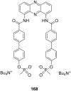
Tweezer 168 for noradrenaline (80b).
Figure 122
Figure 122
Different tripods and heparin (170).
Figure 123
Figure 123
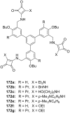
Squaramide based receptors 172.
Figure 124
Figure 124
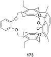
Cage like NH4+ receptor 173 of Kim et al.
Figure 125
Figure 125
Ammonium receptors 174 of Chin et al.
Figure 126
Figure 126
2-Oxazolin-based ammonium receptors 175ad and 176 by Ahn et al.
Figure 127
Figure 127
Racemic guest molecules 177.
Figure 128
Figure 128
Tripods based on a imidazole containing macrocycle (178) and the guest molecules employed in the study (20a, 179a–d).
Figure 129
Figure 129
Ammonium ion receptor 180.
Figure 130
Figure 130
Tetraoxa[3.3.3.3]paracyclophanes 181 and a cyclophanic tetraester (182).
Figure 131
Figure 131
Peptidic bridged paraquat-cyclophane.
Figure 132
Figure 132
Shape-selective noradrenaline host.
Figure 133
Figure 133
Receptor 185 for binding of noradrenaline on surface layers from Schrader et al.
Figure 134
Figure 134
Tetraphosphonate receptor for binding of noradrenaline.
Figure 135
Figure 135
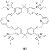
Tetraphosphonate 187 of Schrader and Finocchiaro.
Figure 136
Figure 136
Zinc-Porphyrin ammonium-ion receptors 188 and 189 of Mizutani et al.
Figure 137
Figure 137
Zinc porphyrin receptor 190.
Figure 138
Figure 138
Zinc porphyrin receptors 191 capable of amino acid binding.
Figure 139
Figure 139
Zinc-porphyrins with amino acid side chains for stereoinduction.
Figure 140
Figure 140
Bis-zinc-bis-porphyrin based on Tröger’s base 193.
Figure 141
Figure 141
BINAP-zinc-prophyrin derivative 194 and it’s guests.
Figure 142
Figure 142
Bisaryl-linked-zinc-porphyrin receptors.
Figure 143
Figure 143
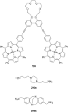
Bis-zinc-porphyrin 199 for diamine recognition and guests.
Figure 144
Figure 144
Bis-zinc-porphyrin crown ether 201.
Figure 145
Figure 145
Bis-zinc-porphyrin 202 for stereodiscrimination (L = large substituent; S = small substituent).
Figure 146
Figure 146
Bis-zinc-porphyrin[3]rotaxane and its copper complex and guests.
Figure 147
Figure 147
Dien-bipyridyl ligand 206 for co-ordination of two metal atoms.
Figure 148
Figure 148
The ligand and corresponding tetradentate co-complex 207 serving as enantioselective receptor for amino acids.
Figure 149
Figure 149
Bis(oxazoline)–copper(II) complex 208 for the recognition of amino acids in aqueous solution.
Figure 150
Figure 150
Zinc-salen-complexes 209 for the recognition tertiary amines.
Figure 151
Figure 151
Bis(oxazoline)–copper(II) 211 for the recognition of amino acids in aqueous solution.
Figure 152
Figure 152
Zn(II)-complex of a C2 terpyridine crown ether.
Figure 153
Figure 153
Displacement assay and receptor for aspartate over glutamate.
Figure 154
Figure 154
Chiral complex 214 for a colorimetric displacement assay for amino acids.
Figure 155
Figure 155
Metal complex receptor 215 with tripeptide side arms.
Figure 156
Figure 156
A sandwich complex 216 and its displaceable dye 217.
Figure 157
Figure 157
Lanthanide complexes 218220 for amino acid recognition.
Figure 158
Figure 158
Nonactin (221), valinomycin (222) and vancomycin (223).
Figure 159
Figure 159
Monesin (224a) and a chiral analogue for enantiodiscrimination of ammonium guests (224b).
Figure 160
Figure 160
Chiral podands (226) compared to pentaglyme-dimethylether (225) and 18-crown-6 (4).
Figure 161
Figure 161
Lasalocid A (228).
Figure 162
Figure 162
Lasalocid derivatives (230) of Sessler et al.
Figure 163
Figure 163
The Coporphyrin I tetraanion (231).
Figure 164
Figure 164
Linear and cyclic peptides for ammonium ion recognition.
Figure 165
Figure 165
Cyclic and bicyclic depsipeptides for ammonium ion recognition.
Figure 166
Figure 166
α-Cyclodextrin (136a) and novocaine (236).
Figure 167
Figure 167
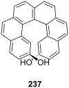
Helical diol receptor 237 by Reetz and Sostmann.
Figure 168
Figure 168
Ammonium binding spherand by Cram et al. (238a) and the cyclic[6]metaphenylacetylene 238b in comparison.
Figure 169
Figure 169
Receptor for peptide backbone and ammonium binding (239).
Figure 170
Figure 170
Anion sensor principle with 3-hydroxy-2-naphthanilide of Jiang et al.
Figure 171
Figure 171
7-bromo-3-hydroxy-N-(2-hydroxyphenyl)naphthalene 2-carboxamide (241) and its amine binding.
Figure 172
Figure 172
Naturally occurring catechins with affinity to quaternary ammonium ions.
Figure 173
Figure 173
Spiropyran (244) and merocyanine form (244a) of the amino acid receptors of Fuji et al.
Figure 174
Figure 174
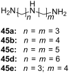
Coumarin aldehyde (245) and its iminium species with amino acid bound (245a) by Glass et al.
Figure 175
Figure 175
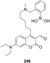
Coumarin aldehyde appended with boronic acid.
Figure 176
Figure 176
Quinolone aldehyde dimers by Glass et al.
Figure 177
Figure 177
Chromogenic ammonium ion receptors with trifluoroacetophenone recognition motifs.
Figure 178
Figure 178
Chromogenic ammonium ion receptor with trifluoroacetophenone recognition motif bound on different matrices.

Similar articles

Cited by

References

    1. Braman S S. N Engl Reg Allergy Proc. 1987;8(2):116–120. doi: 10.2500/108854187778994446. - DOI - PubMed
    1. Idzko M, la Sala A, Ferrari D. J Allergy Clin Immunol. 2002;109(5):839–846. doi: 10.1067/mai.2002.124044. - DOI - PubMed
    1. Yanai K, Tashiro M. Pharmacol Ther. 2007;113(1):1–15. doi: 10.1016/j.pharmthera.2006.06.008. - DOI - PubMed
    1. Ito C. Drug News Perspect. 2004;17(6):383–387. doi: 10.1358/dnp.2004.17.6.829029. - DOI - PubMed
    1. Hubbard B K, Walsh C T. Angew Chem, Int Ed. 2003;42:730–765. doi: 10.1002/anie.200390202. - DOI - PubMed

LinkOut - more resources